The Intraoperative Diagnosis and Management of CSF Leaks in Endoscopic Sinus and Skull Base Surgery. New Paradigm: Topical Fluorescein
Zeina Korban*, Nader Al Souky, Racha Abi Melhem and Usamah Hadi
American University of Beirut, PO Box: 11-0236, Riad El Solh, 1107 2020, Beirut, Lebanon
*Address for Correspondence: Zeina Korban, American University of Beirut, Riad El Solh, 1107 2020, Beirut, Lebanon, Tel: +961-373-7264; E-mail: zk27@aub.edu.lb
Submitted: 08 April 2019; Approved: 22 May 2019; Published: 28 May 2019
Citation this article: Korban Z, Souky NA, Melhem RA, Hadi U. The Intraoperative Diagnosis and Management of CSF Leaks in Endoscopic Sinus and Skull Base Surgery. New Paradigm: Topical Fluorescein. Int J Rhinol Otological. 2019;2(1): 001-004.
Copyright: © 2019 Korban Z, et al. This is an open access article distributed under the Creative Commons Attribution License, which permits unrestricted use, distribution, and reproduction in any medium, provided the original work is properly cited
Keywords: Cerebrospinal fluid leaks; Endoscopic sinus surgery; Topical fluorescein
Download Fulltext PDF
Endoscopic techniques have been widely adopted in sinus and skull base surgery replacing the traditional intracranial/extracranial approaches. However, one of the most common complication is an iatrogenic Cerebrospinal Fluid (CSF) leak, which places the patient at risk for meningitis, pneumocephalus and brain herniation. The instant intraoperative CSF leak diagnosis and consequent repair is crucial to avoid these complications. Traditionally, intrathecal fluorescein injection was utilized to aid in the diagnosis. However, this technique is not FDA approved and encompasses major drawbacks (seizure, lower extremity paresis, cranial nerve palsies and patient discomfort). The purpose of this case series is to introduce a new diagnostic paradigm using topical fluorescein strips “FLUORETS”. It has shown promising prospects due to its safety, accuracy and sensitivity in identifying CSF leaks.
Introduction
Endoscopic techniques represent a major advancement in sinus and skull base surgery achieving higher success rates and lower morbidity than the traditional intracranial and extracranial approaches utilized previously. Iatrogenic CSF leaks can be encountered during these Endonasal approaches. Intraoperative CSF leakage is reported to occur between 5-20% of cases [1]. Most commonly, the skull base defects are encountered at the ethmoid roof, cribriform plate and sphenoid sinus. At times, these defects can be missed during the course of the operation and this can be attributed by the defect size and CSF flow rate.
Accurate diagnosis is crucial to avoid the devastating consequences that can ensue ranging from meningitis to brain herniation and persistence of the CSF leak.
While postoperative diagnosis of CSF leaks is well established in the literature and accurate most of the time, intraoperative diagnosis remains a challenging issue.
Various methods have been utilized to accurately diagnosis and prevent CSF leaks intraoperatively. Successful identification of an accidental breach of the skull base relies on several parameters.
Emphasis begins on the significance of preoperative imaging that can raise the surgeon’s index of suspicion for potential intraoperative accidents.
In addition, the presence of intranasal polyposis and revision surgery increase the likelihood of complications. Accurate knowledge of the anatomy and level of expertise of the surgeon are mandatory assets for safe and successful navigation through the sinus labyrinth with satisfactory outcomes.
Multiple mechanisms have been described in the literature pertaining to intraoperative diagnosis of CSF leaks. Traditionally, Intrathecal Fluorescein (ITF) [2] injection was the most commonly used method. However, this technique was associated with major transient side effects including seizures, cranial nerve palsies and lower extremity paresis [3,4]. In addition, this technique is uncomfortable to the patient and therefore can be refuted by many [5] Intraoperatively, CSF is translucent and can be obscured by the presence of blood and mucosal secretions. Localizing defects intraoperatively begins with direct visualization using the endoscope, assisted by the surgeon’s index of suspicion. Utilizing Valsalva maneuvers has been described as a provocative measure to help precipitate CSF flow through breached dura during venous engorgement. However; these maneuvers are not always rewarding leaving the surgeon perplexed regarding the diagnosis and management.
We hereby introduce the concept of utilizing topical intranasal fluorescein strips “Fluorets” as a safe, accurate and sensitive tool that assists in localizing defects and identifying suspicious intraoperative CSF leaks in addition to confirming the absence of a CSF leak after a successful repair.
In this technique, a topical intranasal fluorescein strip is applied intraoperatively. A change in the color of the secretions from yellow/brown to green indicates the presence of CSF and allows tracing back the source of the leak. Following the diagnosis, endoscopic repair will be carried out. If there is no visual CSF leak even with a Valsalva manoeuver, the technique can be repeated to confirm a water tight closure.
Case Report
We have resorted to the utilization of topical fluorescein strips on ten occasions (4 ethmoid roof defects and 6 sphenoid sinus defects) in our institution for the diagnosis of CSF leaks. It has accurately detected CSF leaks in both endoscopic sinus surgery and transphenoidal pituitary surgery. Three cases will be used for illustration.
Method
All cases involved utilization of a topical ophthalmologic fluorescein strip (Flouret) during endoscopic sinonasal surgery and endoscopic transphenoidal resection of pituitary adenomas and laid at the site of the expected defect. CSF was confirmed by green discoloration at the site of the defect. Multilayered repair then ensued after confirmation.
Case 1
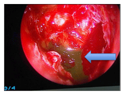
We hereby present the case of a 55-year-old gentleman, BMI (Body Mass Index) of 25, with a history that is remarkable for endoscopic sinus surgery ten years prior to presenting to our institution. Patient was diagnosed and treated for meningitis on two occasions. Preoperative work-up, including CT with metrizamide, failed to demonstrate the defect site. Patient did not consent for intrathecal fluorescein. Intraoperatively, under direct visualization, with the assistance of Valsalva maneuvers, no defect could be localized. We then utilized a topical fluorescein strip through the nasal cavity and laid it against the ethmoid roof after stripping the mucosa. Green discoloration was encountered at the posterior aspect of the ethmoid in proximity to the anterior face of the sphenoid sinus where a defect was then identified (Figure 1). Consequently adequate repair, in a multilayer fashion, was performed utilizing temporalis fascia grafts supplemented with fibrin glue and absorbable surgical followed by merocel packs. Postoperative care was smooth with no evidence of recurrence at one-year follow up.
Case 2
We present another case of a 30-year-old female, BMI of 20 diagnosed with a craniopharyngioma that failed initial resection, presented to our institute for endoscopic endonasal transphenoidal surgery.
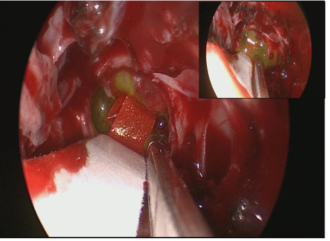
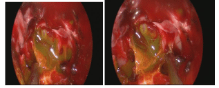
Intraoperatively, following resection, the sterile strip technique of fluorescein was applied at the lateral face of the sphenoid sinus. A pool of green discoloration was seen indicating the defect site (Figure 2, 3a, 3b). Repair was attempted using a multilayer technique. Abdominal fat was also harvested and used for the repair. At the end of the repair where no CSF was directly visualized, application of a fluoret strip revealed a persistent leak. Further repair using mucosal grafts was performed; following which another strip was performed demonstrating a successful repair with no discoloration.
Case 3
A 47-year-old lady, with an elevated BMI of 30, presented with spontaneous clear rhinorrhea. She denies any history of trauma, or sinonasal surgery. Nasal endoscopy was done and CSF analysis with Beta 2 transferrin confirmed CSF.
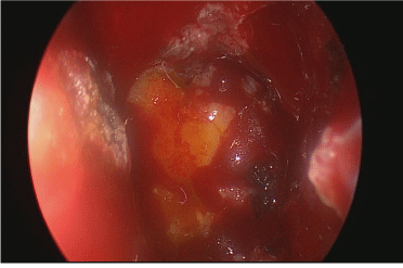
Following repair of the defect at the cribriform plate using a multilayer technique supplemented by a free septal mucosal graft, a fluoret strip was used to document complete cessation of CSF leak and hence secure repair of the defect (Figure 4).
Discussion
Galen first described CSF rhinorrhea in the second century AD [6]. The first repair of a CSF leak did not occur until 1700 years later, when Dandy performed an open based craniotomy procedure using a fascia lata graft for transcranial skull reconstruction [7]. In 1948, Dohlman was the first to utilize the extracranial approach via an external ethmoidectomy [8] making it the standard of care with success rates as low as 60% [9]. Endoscopic repair of CSF leaks did not occur until 1981when it was first described by Wigand [10].
The endoscopic approach is a safe and effective procedure, which has grown to be the standard of care for most anterior skull base defects with success rates approaching 90% [11]. Principles mirror those of open repair with lower operative morbidity and higher accuracy. The most frequent inciting factor for CSF leaks is accidental trauma (44%) followed by surgical trauma (29%) and tumors, whether benign or malignant (22%) [12]. Congenital and spontaneous sources have also been described in literature [12].
Iatrogenic CSF leaks are attributed to transphenoidal resection of pituitary tumors (15%), and functional endoscopic sinus surgery (3%) [1]. Most can be missed, and on other occasions, the leak is suspected but cannot be proven. Hence, inadvertent or no repair may be performed.
Accurate diagnosis and localization of defects can be challenging even for the most experienced surgeon. Localizing the site of origin is fundamental and plays a role in the surgical approach. Iatrogenic CSF leaks most commonly arise at the ethmoid roof (35%), followed by the cribriform plate (27%) and the sphenoid sinus (19%) [13]. Otolaryngologists’ experience with CSF leaks is primarily composed of repair post sinus surgery or transphenoidal surgery.
Intraoperative CSF leaks are reported in 15-30%. Postoperative complications of CSF leaks and inadequate repair include tension pneumocephalus and meningitis [2]. Ozturk et al. found that 46% and 36% of patients presenting with CSF leakage had meningitis or recurrent meningitis respectively [5].
At present, some authors advocate the use of lumbar drains alone or in adjunct with intrathecal fluorescein to assist in graft placement and localize fistulas. [1] Intrathecal fluorescein can cause potential complications and is not FDA approved. This has limited its clinical use.
Topical strip intranasal fluorescein introduces a new, minimally invasive, simple and convenient method for localizing CSF fistulas. This technique was recently utilized in our institute in all our cases of suspected CSF leaks with a high success rate.
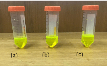
This consists of applying fluorescein strips (Fluorets) that are sterile and used by ophthalmologists to stain corneal ulcers. Fluorescein strip gives a yellow color when it gets in contact with water, saline and nasal secretions (Figure 5). A change to green color indicates presence of CSF.
The concept of topical fluorescein was first utilized by Jones whereby he achieved 100% accuracy in localizing a CSF leak in 3 patients with no adverse side effects. He introduced the concept of cotton pledgets soaked in fluorescein [14]. However; this technique has its disadvantages in terms of resources, standardization of concentrates and intranasal contact time. Saafan was also successful in achieving 100 % accuracy used topical intranasal 5% fluorescein for preoperative diagnosis and intraoperative localization of the site of the leak [15].
Topical nasal fluorescein is an easy, sensitive, safe and highly accurate tool in the intraoperative localization of the site and extent of CSF fistulas. In our hands, it has proven to be effective in diagnosing the site of CSF leak. It has also been useful in documenting complete seal after a presumed successful repair. It bypasses the adverse side effects associated with the use of a lumbar drain and intrathecal fluorescein and should be sought as a noninvasive alternative and a principle tool in diagnosis. Topical intranasal fluorescein represents an alternative method for skull base repair after tumor resection avoiding the need of a lumbar drain and the use of intrathecal fluorescein. In addition; the use of intrathecal fluorescein may be refuted on occasions by patients after the risk is explained. The use of the fluorescein strip technique avoids these consequential issues and is widely accepted by patients in our institution. However, one drawback to this technique is its inability to detect multiple defects of CSF leakage while intrathecal fluorescein injection does.
Conclusion
Repair of skull base defects using minimally invasive and endoscopic techniques continues to evolve. A comprehensive understanding of the anatomy, physiology, diagnosis and surgical approaches is needed to ensure proper treatment. Diagnostic measures play a crucial role in preventing adverse sequelae. We have achieved success in identifying leak sites using our technique of topical fluorescein strips (Hadi-Korban Technique) that has proven to be effective in diagnosing the site of CSF leak and documenting complete seal after a presumed successful repair. It has introduced a new paradigm in diagnosis and management.
- Ruggeri AG, Cappelletti M, Giovannetti F, Priore P, Pichierri A, Delfini R. Proposal of standardization of closure techniques after endoscopic pituitary and skull base surgery based on postoperative cerebrospinal fluid leak risk classification. J Craniofac Surg. 2019. https://bit.ly/2Hu8zQA
- Raza SM, Banu MA, Donaldson A, Patel KS, Anand VK, Schwartz TH. Sensitivity and specificity of intrathecal fluorescein and white light excitation for detecting intraoperative cerebrospinal fluid leak in endoscopic skull base surgery: A prospective study. J Neurosurg. 2016; 124: 621-626. https://bit.ly/2LZx1Oh
- Keerl R, Weber RK, Draf W, Wienke A, Schaefer SD: Use of sodium fluorescein solution for detection of cerebrospinal fluid fistulas: an analysis of 420 administrations and reported complications in Europe and the United States. Laryngoscope. 2004; 114: 266-272. https://bit.ly/2HFqiDt
- Moseley JI, Carton CA, Stern WE: Spectrum of complications in the use of intrathecal fluorescein. J Neurosurg. 1978; 48: 765-767. https://bit.ly/30ySgJU
- Ozturk K, Karabagli H, Bulut S, Egilmez M, Duran M. Is the use of topical fluorescein helpful for management of CSF leakage?. Laryngoscope. 2012; 122: 1215-1218. https://bit.ly/2Qh9g2r
- Wax MK, Ramadan HH, Ortiz O, Wetmore SJ. Contemporary management of cerebrospinal fluid rhinorrhea. Otolaryngol Head Neck Surg. 1997; 116: 442-449. https://bit.ly/2wd4kCK
- White DR, Dubin MG, Senior BA. Endoscopic Repair of cerebrospinal fluid leaks after Neurosurgical Procedures; Am J Otolaryngol. 2003; 24: 213-216. https://bit.ly/2HLeboD
- Dohlman G. Spontaneous cerebrospinal fluid rhinorrhea. Acta Otolarygol (Stockh). 1948; 67: 20-23.
- Senior BA, Jafri K, Benninger M: Safety and efficacy of endoscopic repair of CSF leaks: A survey of the members of the American Rhinologic Society. Am J Rhinol. 2001; 15: 21-25. https://bit.ly/2Hv82Ot
- Wigand ME. Transnasal ethmoidectomy under endoscopic control. Rhinology. 1981; 19: 7-15. https://bit.ly/2wd0LfI
- Hegazy HM, Carrau RL, Snyderman CH, Kassam A, Zweig J. Transnasal endoscopic repair of cerebrospinal fluid rhinorrhea: A meta-analysis. Laryngoscope. 2000; 110: 1166-1172. https://bit.ly/2QgsOnJ
- Lopatin AS, Kapitanov DN, Potapov AA. Endonasal endoscopic repair of spontaneous cerebrospinal fluid leaks. Arch Otolaryngol Head Neck Surg. 2003; 129: 859-863. https://bit.ly/30Bq5tF
- Locatelli D, Rampa F, Acchiardi I, Bignami M, De Bernardi F, Castelnuovo P. Endoscopic endonasal approaches for repair of cerebrospinal fluid leaks: nine-year experience. Neurosurgery. 2006; 58: 246-256. https://bit.ly/2HsEMYs
- Jones ME, Reino T, Gnoy A, Guillory S, Wackym P, Lawson W. Identification of intranasal cerebrospinal fluid leaks by topical application with fluorescein dye. Am J Rhinol. 2000; 14: 93-96. https://bit.ly/2M8TjgC
- Saafan ME, Ragab SM, Albirmawy OA. Topical intranasal fluorescein: the missing partner in algorithms of cerebrospinal fluid fistula detection. Laryngoscope. 2006; 116: 1158-1161. https://bit.ly/2YzgIJc

Sign up for Article Alerts