Sub-Prosthetic Total Abdominal Recurrent Herniation Following Incisional Hernia Repair with a 30x30 Cm Onlay Mesh: Case Report
Hakan Kulacoglu1* and Haydar Celasin2
1Ankara Hernia Center, Ankara, Turkey2Lokman Hekim Akay Hastanesi, Ankara, Turkey
*Address for Correspondence: Hakan Kulacoglu, Ankara Hernia Center, Cukurambar mh. Budapeste Cd. 33/A 06530 Ankara Tel: +905-055-254-000; Fax: +903-122-200-090; E-mail: [email protected]; [email protected]
Submitted: 05 February 2018; Approved: 26 March 2018; Published: 29 March 2018
Citation this article: Kulacoglu H, Celasin H. Sub-Prosthetic Total Abdominal Recurrent Herniation Following Incisional Hernia Repair with a 30x30 Cm Onlay Mesh: Case Report. Open J Surg. 2018;2(1): 011-014.
Copyright: © 2018 Kulacoglu H, et al. This is an open access article distributed under the Creative Commons Attribution License, which permits unrestricted use, distribution, and reproduction in any medium, provided the original work is properly cited
Download Fulltext PDF
Recurrence following incisional hernia repair is a common problem even after mesh use. We present a 42-year-old woman with a giant re-recurrent incisional hernia. Hernia recurred despite a 30x30 cm onlay mesh. The whole area of the mesh was covered inside with peritoneum till both axillary lines. The patient was treated with sublay mesh repair following transversus abdominis muscle release.
Introduction
The incidence of incisional hernia following laparotomies can be higher than 15% (Nakayama). Risk increases when the patient is obese [1,2]. Recurrence rates after incisional hernia repairs are also higher in comparison with groin hernia repairs [3]. Ten-year recurrence rates have been reported as 60% for suture repair and 30% for mesh augmentation [4]. Re-repairs results in two-fold high failure rates than primary repairs [5]. Herein, a case of re-recurrent incisional hernia despite a prosthetic repair with 30 x 30 cm mesh is presented.
Case Presentation
A 42-year-old female patient was admitted with a complaint of abdominal enlargement, pain and limitation of physical activity. She was 165 cm in height and weighed 92 kilograms. Her body mass index was 33.8 (Grade 1 Obesity). In medical history, she had no systemic disorders, but well-controlled hypothyroidism. She underwent 4 abdominal operations within a 4-year period. The first surgery was an abdominoplasty following her second pregnancy. The second one was abdominal hysterectomy via a Pfannensteil incision. Two years and 4 months later she developed an incisional hernia on gynecologic surgery site. An open onlay mesh repair was performed but the hernia recurred after three months. The last operation was one year ago, it was again an open mesh repair again by using a 30x30 cm polypropylene mesh in onlay position. Nevertheless, she started feeling uncomfortable after one year. Several physical examinations by different surgeons did not detect a recurrence despite a luxation on the abdominal wall. Besides, abdominal ultrasound revealed a luxated prosthetic material over a thinned but intact abdominal wall.
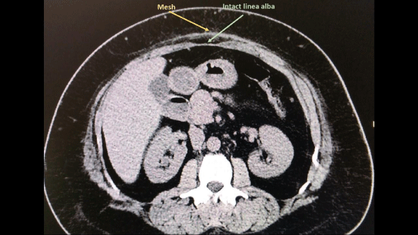
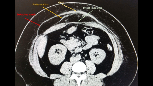
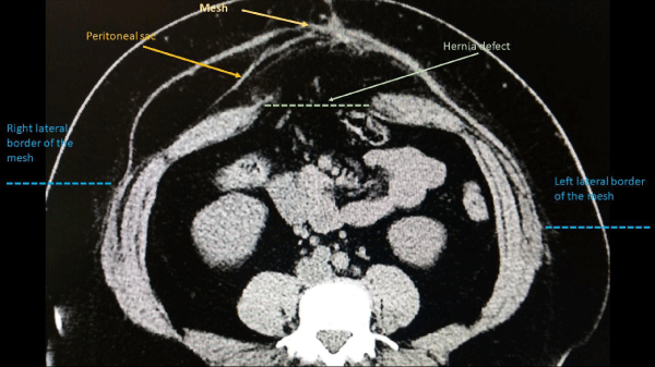
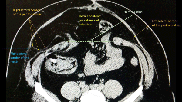
Her complaints got worse with increasing abdominal girth and she admitted for another opinion finally. In physical examination, an apparent luxation was observed in inspection. When Valsalva maneuver was applied herniation of intestinal loops through a large defect became palpable. Computed Tomography (CT) showed a very large incisional hernia underneath an intact mesh. Mesh covered whole abdominal wall, no hernia sac was detected beyond the boundaries of the mesh. The linea alba was wide (diastasis recti) but intact in subxiphoid/epigastric region and covered with mesh (Figure 1). A peritoneal sac became visible at the supraumbilical region while the linea alba is still intact (Figure 2). Subsequently, the linea alba was detached and the hernia content was seen below the umbilical level (Figure 3). Eventually, the limits of peritoneal sacs reached over lateral muscle group on both sides (Figure 4). The hernia defect sizes were 15 cm in width and length.
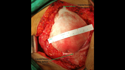
After discussing with the patient of the potential complications and benefits of a new surgical repair and the risks of waiting a re-operation was scheduled. The planned procedure was sublay mesh placement following mesh extraction and TAR (transversus abdominis muscle release). A midline incision was made. Skin flap dissection was advanced towards lateral, superior and inferior borders of the mesh. In accordance with the CT findings no peritoneal sac was seen at the borders of the mesh. The mesh was divided on midline at its uppermost border where no herniation had been observed in CT study. There were almost no intestinal adhesions to the mesh and into the peritoneal sac. The whole area of the mesh was covered inside with peritoneum till both axillary lines (Figure 5). The mesh and the redundancy of peritoneal sac were extracted, and the borders of the rectus muscles were revealed. There were also numerous thick polyester sutures over the anterior rectus sheath possibly put for fixing diastasis recti. These sutures were removed. The defect size was 20x15 cm (Figure 6). Retro muscular dissection was commenced and a large room was prepared for the new mesh with TAR. Posterior sheath and peritoneal flaps was closed with 2/0 polydioxanone. A 30x30 cm polypropylene mesh was placed with transfascial polydioxanone sutures. Anterior sheath was sutured with 0 polydioxanone. Two suctions drains were placed on either side.
The patient tolerated oral food in the following morning. She responded well to oral analgesics and was discharged with recommendation of weight loss. Follow-up examinations were scheduled for every other day. No wound complications were recorded, and the drains were removed on 15th day. She lost 5 kilograms within the first month.
Discussion
Recurrence following incisional hernia repair is a common problem even after mesh use. Prosthetic materials decrease failure rates in hernia surgery, however it is illogical to think that they perform a magical task. Meshes promote inflammatory and fibroblastic reactions after implantation; tissue ingrowth, integration and fibrosis eventually render the repair strong. It is recommended that the size of the mesh provide at least 5 cm overlap towards all directions of the repair line. Mesh shrinkage is a well-known problem, and it has been shown that there is a significant correlation between tissue ingrowth force and mesh size [6]. Indeed, Conze et al reported that recurrences after median incisional hernia mesh repair manifested at the margin of the enclosed mesh [7]. However, overlapping with large meshes cannot always prevent recurrence. When suture line is disrupted because of either poor surgical technique or progressively increased abdominal pressure due to weight gain or some other reasons mesh will not sufficiently support the repair line. A peritoneal protrusion comes up and it advances to a true hernia recurrence by time. Recurrence is easier when a new linea alba is not created and the mesh is used for bridging.
In the present case a 30x30 cm mesh had been used for re-repair. Nevertheless, re-recurrence occurred after a relatively short while. The reason for the recurrence was probably a suture line disruption because of obesity. It is also possible that previous diastasis recti contributed the fascial tears through needle holes. Peritoneal protrusion advanced especially to both lateral directions and a hernia sac which is almost equal to the mesh size was formed beneath the mesh.
In fact, recurrence after incisional hernia repair is not only more frequent than inguinal hernia repairs but also appears earlier in comparison with them. Köckerling et al reported that one third of all recurrences after incisional hernia repairs occurred in the first postoperative year, and two thirds within two to three years [8]. Re-repair for a recurrence despite a previous mesh repair is a real surgical challenge and re-repairs may result in two-fold high failure rates than primary repairs [5,7], therefore surgical experience is of importance [9]. Several studies revealed that sublay (retro rectus) mesh placement provide better results including low recurrence rates [10-12]. This technique supports abdominal wall against median disruption and seems to be the proper choice for patients with recurrent hernias and high body mass index like the present case.
Correct diagnosis of incisional hernias may be difficult especially in obese patients. Baucom et al reported that surgeon’s physical examination is inferior to CT for detection of incisional hernia; it fails to detect approximately one third of hernias in obese patients [13]. It has also been shown that CT is very helpful in the detection of recurrence after incisional hernia repair [14]. Physical examination may fail especially in cases with an intact mesh over a recurrent hernia mass. Ultrasound is a user dependent diagnostic tool and may also miss the recurrence in some cases, although some researchers claimed that dynamic abdominal ultrasound is a good alternative to CT [15].
In the present case several physical examinations could not correctly diagnosed the recurrence. The reason was probably intact layer of mesh which pretending a healthy fascial layer over the hernia mass. Ultrasound examination also missed the recurrence because of the same condition. Mesh on the midline may have mimicked diastasis recti. Nevertheless, a simple Valsalva maneuver could have revealed the herniation and the borders of the fascial defect.
Conclusion
Recurrent herniation beneath a prosthetic material is possibly not uncommon. However, a subprosthetic herniation toward almost the total area of the mesh is probably very rare. Large onlay mesh is not a guarantee for durability of the repair. The integrity of the suture closure line is important, and a recurrence eventually inevitable when a disruption develops. Sublay mesh technique should be the choice especially in obese patients. Physical examination and even ultrasound is insufficient in certain cases and the diagnosis should be confirmed by computed tomography.
- Nakayama M, Yoshimatsu K, Yokomizo H, Yano Y, Okayama S, Satake M, et al. Incidence and risk factors for incisional hernia after open surgery for colorectal cancer. Hepatogastroenterology. 2014; 61: 1220-1223. https://goo.gl/mGMS93
- Walming S, Angenete E, Block M, Bock D, Gessler B, Haglind E. Retrospective review of risk factors for surgical wound dehiscence and incisional hernia. BMC Surg. 2017; 17: 19. https://goo.gl/1V8JVb
- Sanders DL, Kingsnorth AN, Windsor AC. Is there a role for hernia subspecialists? Or is this a step too far? Hernia. 2016; 20: 637-640. https://goo.gl/PmnsUJ
- Burger JW, Luijendijk RW, Hop WC, Halm JA, Verdaasdonk EG, Jeekel J. Long-term follow-up of a randomized controlled trial of suture versus mesh repair of incisional hernia. Ann Surg. 2004; 240: 578-583; discussion 583-5. https://goo.gl/cxMdqX
- Pereira JA, Lopez Cano M, Hernández-Granados P, Feliu X; en representación del grupo EVEREG. Initial results of the National Registry of Incisional Hernia. Cir Esp. 2016; 94: 595-602. https://goo.gl/3q2Kjg
- Gonzalez R, Fugate K, McClusky D 3rd, Ritter EM, Lederman A, Dillehay D, et al. Relationship between tissue ingrowth and mesh contraction. World J Surg. 2005; 29: 1038-1043. https://goo.gl/1GgXxm
- Conze J, Krones CJ, Schumpelick V, Klinge U. Incisional hernia: challenge of re-operations after mesh repair. Langenbecks Arch Surg. 2007; 392: 453-457. https://goo.gl/MVMgQ5
- Kockerling F, Koch A, Lorenz R, Schug-Pass C, Stechemesser B, Reinpold W. How Long Do We Need to Follow-Up Our Hernia Patients to Find the Real Recurrence Rate? Front Surg. 2015; 2: 24. https://goo.gl/CicQ47
- Aquina CT, Kelly KN, Probst CP, Iannuzzi JC, Noyes K, Langstein HN, et al. Surgeon volume plays a significant role in outcomes and cost following open incisional hernia repair. J Gastrointest Surg. 2015; 19: 100-110. https://goo.gl/i3PAZ1
- Albino FP, Patel KM, Nahabedian MY, Sosin M, Attinger CE, Bhanot P. Does mesh location matter in abdominal wall reconstruction? A systematic review of the literature and a summary of recommendations. Plast Reconstr Surg. 2013; 132: 1295-1304. https://goo.gl/35rYdn
- Holihan JL, Nguyen DH, Nguyen MT, Mo J, Kao LS, Liang MK. Mesh location in open ventral hernia repair: a systematic review and network meta-analysis. World J Surg. 2016; 40: 89-99. https://goo.gl/SKgmVL
- Timmermans L, de Goede B, van Dijk SM, Kleinrensink GJ, Jeekel J, Lange JF. Meta-analysis of sublay versus onlay mesh repair in incisional hernia surgery. Am J Surg. 2014; 207: 980-988. https://goo.gl/SLRUXJ
- Baucom RB, Beck WC, Holzman MD, Sharp KW, Nealon WH, Poulose BK. Retrospective evaluation of surgeon physical examination for detection of incisional hernias. J Am Coll Surg. 2014; 218: 363-366. https://goo.gl/zhYYhf
- Gutiérrez de la Peña C, Vargas Romero J, Diéguez García JA. The value of CT diagnosis of hernia recurrence after prosthetic repair of ventral incisional hernias. Eur Radiol. 2001; 11: 1161-1164. https://goo.gl/NkoB7V
- Beck WC, Holzman MD, Sharp KW, Nealon WH, Dupont WD, Poulose BK. Comparative effectiveness of dynamic abdominal sonography for hernia vs computed tomography in the diagnosis of incisional hernia. J Am Coll Surg. 2013; 216: 447-453. https://goo.gl/ESEHwJ


Sign up for Article Alerts