Surgical Treatment of Hepatic Hydatid via Low-Traumatic Approach (the Experience of the Peace-support Mission in Afghanistan)
Ievgen Tsema1* and Vladimir Negoduyko2
1Surgical Department #4 , Bogomolets National Medical University, Kyiv, Ukraine
2Department of Abdominal Surgery, Military-Medical Clinical Center of North Region, Kharkiv, Ukraine
*Address for Correspondence: Ievgen Tsema, Surgical Department #4 , Bogomolets National Medical University, Shovkovychna 39/1, 01601 Kyiv, Ukraine, Tel: +38 (063) 731-59-95; E-mail: hemorrhoid@ukr.net
Submitted: 06 October 2017; Approved: 20 October 2017; Published: 21 October 2017
Citation this article: Tsema I, Negoduyko V. Surgical Treatment of Hepatic Hydatid via Low-Traumatic Approach (the Experience of the Peace-support Mission in Afghanistan). Open J Surg. 2017;1(1): 015-019.
Copyright: © 2017 Tsema I, et al. This is an open access article distributed under the Creative Commons Attribution License, which permits unrestricted use, distribution, and reproduction in any medium, provided the original work is properly cited
Keywords: Hepatic hydatid; Surgical treatment; Low-traumatic approach
Download Fulltext PDF
The aim of the study was to estimate the results of using the low-traumatic approach in the surgical treatment of patients with large hepatic hydatids.
Materials and methods: The results of the surgical treatment of 81 patients with large hydatid cysts in the Islamic Republic of Afghanistan have been analyzed. The patients were divided into 2 groups. The main group consisted of 40 patients, which underwent hydatidectomy via low-traumatic approach. The control group consisted of 41 patients, which underwent hydatidectomy via traditional open laparotomy. Both groups of the patients were comparable by sex, age, and stage and cyst localization.
Results: It was defined that 76 (93.8%) patients had large and giant hepatic hydatids. The duration of the disease manifestation in the patients varied from 3 months to 12 years. 63 (77.8%) patients had foreign body feeling in the abdominal cavity and constant or intermittent pain in the right hypochondrium. 18 (22.2%) patients had palpable neoplasm but they didn’t have any visible abdominal deformations. Hydatidectomy using the low-traumatic approach was possible in 38 (95.0%) patients of the main group. The usage of the proposed approach allowed to significant improvement in the life quality characteristics of the patient in the early postoperative period, such as a total duration of the in-patient treatment, a duration of the in-patient treatment after surgery, a duration of postoperative pain syndrome requiring the use of an analgesic, a duration of the intestinal peristalsis recovery, a duration of the infusion therapy after the surgery, a duration of the bed rest. Moreover, the use of low-traumatic approach allowed to significant reduction of the time of returning to a habitual physical activity and to essential decrease of a duration of discomfort feeling in the wound area.
Conclusion: Usage of the low-traumatic surgical approach is a reliable and a safe method of treatment of the patients with large and giant hepatic hydatids, including ones complicated by suppuration. Furthermore, the proposed approach can be useful for the hepatic hydatid treatment in the endemic developing countries, where there is no access to video image endoscopy surgical equipment.
Introduction
The existence of endemic areas of the hydatid disease in the developing countries enables to obtain valuable experience of the treatment for this pathology. It becomes possible due to medical care delivery to the local population in the peace-support and humanitarian international missions [1-4]. There are some difficulties in the surgical treatment of hydatid cysts of the liver at present time. Firstly, the traditional methods, which are used for the surgical treatment of large hydatid cysts of the liver, require a quite traumatic surgical approach [5]. The use of laparoscopic methods is limited both by the size of hydatid cysts and by the low availability of the minimally invasive technologies in the developing countries [5-7]. One of directions in resolving of these problems is the implementation of the surgical interventions with using of the low-traumatic approach. First of all, it allows to reduce the operational trauma in cases of a large size hepatic hydatids removal [4].
Materials and Methods
The outcomes of the surgical treatment of 81 patients with hepatic hydatids have been represented in this research. All patients were treated at Surgical Department of Chakcharan Provincial Hospital and Medical Section of Advanced Operative Base “Shield” of Islamic Republic of Afghanistan during the period from 2008 to 2014 year. All the patients were residents of the mountainous regions of Afghanistan. The main group consisted of 40 patients, which underwent hydatidectomy via the low-traumatic approach. The control group consisted of 41 patients, which underwent hydatidectomy via traditional surgical approach. Both groups of the patients were comparable by sex, age (Table 1), stage and cyst localization (reliability of differences between the control and the main group of the patients was < 0.05). The stage of hydatid cyst’s development was evaluated according to Akhmedov’s classification [8]. The classification recommended by the World Health Organization was used to determine cyst’s size [6]. The preoperative ultrasound investigation was performed in all patients with hepatic hydatids. It allowed to define the stage of the cyst’s development and to determine their position in the liver segments. The study results processing was carried out using parametric statistics.
Results
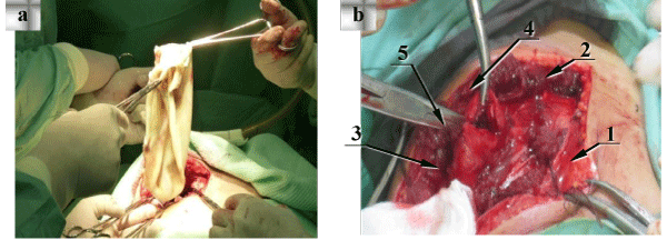
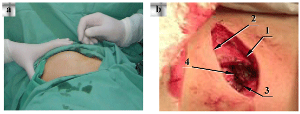
All researched patients had clinical manifestations of the disease. The duration of the disease manifestation in the patients varied from 3 months to 12 years. The reasons for a doctor’s visit were the symptoms of cyst suppuration or a visible outpouching of hepatic hydatid on the anterior abdominal wall. A large hepatic hydatid was diagnosed in 62 (76.5%) patients, a giant cyst – in 14 (17.3%) patients and only 5 (6.2%) patients had a medium sized hepatic cyst. The enlargement of abdomen and the deformation of its anterior wall were the most frequent combination of the clinical symptoms in patients with hydatid disease of the liver. There were foreign body feeling in the abdominal cavity and a constant or intermittent pain in the right hypochondrium in 63 (77.8%) patients. 18 (22.2%) patients had a palpable neoplasm but they didn’t have any visible abdominal deformations. In our practice, we had only one woman with hepatic hydatid who had neither an enlargement of the abdomen nor a palpable neoplasm. This patient had a rupture of hydatid cyst that led to diffuse peritonitis. This case wasn’t included into this study, because the patient had died in the first hours after operation. We used one of three variants of the surgery approach in the patients of the control group: midline laparotomy, Kocher’s incision or anterolateral laparotomy. 5 patients of the control group had hepatic hydatid in the left lobe of the liver. Thus, Kocher’s incision was used in 2 patients which had hepatic hydatid in S1, midline laparotomy was used in 2 patients which had hepatic hydatid in S2, and also midline laparotomy was used in 1 patient who had cysts in S2 and S3 of the liver. According to the algorithm of a surgery approach choice, Kocher’s incision was used in the cases of the hydatid cysts localization in the “front” segments (S5, S6) of the right hepatic lobe; whereas, either anterolateral laparotomy or Kocher’s incision was used in the patients with the localization of hepatic hydatids in the “posterior” segments (S7, S8) of the right hepatic lobe. Generally, Kocher’s incision was used in the cases of the hepatic hydatids localization in S8 – the incision was performed in the area of cyst’s projection on the anterior abdominal wall. The distribution of the patients of the control group with dextral lobe’s hepatic hydatids depending on the chosen surgery approaches is presented in the (Table 2). After performing the surgical approach to a hepatic hydatid, the puncture of the cyst and its decompression (hydatid contents aspiration) have been carried out. For this purpose, the free abdominal cavity was delimited with tampons moistened with 25% sodium sulfate solution. The next step was to remove the chitinous capsule of hepatic hydatid (Figure 1a), after that its remaining fibrous tunic was sutured with the parietal peritoneum (marsupialization of hepatic hydatid, (Figure 1b). Then, cyst’s cavity draining and the wound closure were carried out. A drainage tube was inserted through the incisional wound in 31 (75.6%) cases; beside to the operative wound (through the separate counter opening) – in 6 (14.6%) patients and both through and beside the incisional wound – in 4 (9.8%) patients with multiple hepatic hydatids. The same steps of the operation were performed like in an uncomplicated hydatid cyst in the case of a festered hepatic hydatid (Figure 2). Treatment of the patients of the main group was carried out using the low-traumatic approach for reducing a duration of in-patient treatment and increasing the patients’ life quality after the operation. The low-traumatic approach was conducted via performing of an incision no longer than 6 cm in the place chosen according to the algorithm described above for the patient of the control group (Figure 3b). All patients of both groups had the palpable signs concerning a localization of hepatic hydatid and 62 (76.5%) patients had a visible cyst’s outpouching which indicated the projection of hepatic hydatid on the anterior abdominal wall (Figure 3a). Thereby, a localization of hepatic hydatid was possible to determine in all patients even after the physical examination only. The definite localization of a cyst in the liver parenchyma was determined using ultrasound investigation. The optimal position for a surgical approach was determined depending on the obtained data about cyst’s position relatively to the anterior abdominal wall. Midline mini-laparotomy in the main group was performed in 3 (7.5%) patients with hepatic hydatid of the left liver lobe (2 patients – S2 and 1 case – S1). The distribution of the patients of the main group with dextral lobe’s hepatic hydatids depending on the chosen surgery approach is presented in the Table 3. Thereby, hydatidectomy using the low-traumatic approach was possible in 38 (95.0%) patients of the main group. The analysis of the spent time for surgical intervention showed that increasing of our operating crew experience in the low-traumatic hydatidectomy and developing of our operative technique led to decreasing of an average duration of the operation (Figure 4). It was carried out the comparative analysis of the next main indices of the early postoperative course in the control and the main group of the patients, such as a total duration of the in-patient treatment, a duration of the in-patient treatment after the surgery, a duration of a postoperative pain syndrome requiring the use of analgesics, a duration of a recovery intestinal peristalsis, a duration of an infusion therapy after the surgery, a duration of the bed rest (Table 4). An ultrasound investigation of the liver in the postoperative period showed that in both groups of the patients there was the gradual decrease of the cavity size after hepatic hydatid removal. The obtained data correlated with an average time of drain’s staying, an average term of discomfort feeling in the wound area and an average term of returning to a habitual physical activity of the researched patients (Table 5). Thereby, a surgical intervention performing with usage of the low-traumatic approach didn’t lead to a deterioration of the long-term results of the residual cavity drainage and didn’t prolong a time of drain’s staying. Furthermore, using the low-traumatic surgical approach allowed to reduce significantly the time of returning to a habitual physical activity and to decrease essentially the duration of discomfort feeling in the wound area.
Discussion
The introduction of modern technologies in a surgical treatment of patients allowed to minimize the traumatism of extensive surgical interventions (such as hemicolectomy, pancreatoduodenal resection, gastrectomy), including echinococcectomy [2,3,9,10]. The traumatic nature of the surgery interventions can be significantly reduced by using laparoscopic methods of the surgical treatment, but their wide application is constrained by the high probability of cyst rupturing and its contents disseminating over the abdominal cavity [10-12]. Early publications about the results of the laparoscopic treatment of hepatic hydatid reported about a high risk of intra-abdominal complications such as a dissemination of parasites, an injury of large vessels and bile ducts, a formation of bile fistulas [2,3]. The 1st, 2nd, 3rd, 5th and 6th hepatic segments are considered as the most safe for performing of the laparoscopic operations. Whereas, performing of the laparoscopic intervention is considered as a dangerous procedure due to poor visualization of an affected site of the liver in the cases of hydatid cyst localization in 4th, 7th and 8th hepatic segments [11,12]. In addition, the use of the laparoscopic surgery is contraindicative in cases of secondary infection of hydatid cyst [2,4,7]. Later research works showed a higher effectiveness of the laparoscopic techniques. However, the laparoscopic interventions still continue to be disputable for using of video image endoscopy in patients with large / giant hepatic hydatid or suppurated cyst [3,5,7,9]. Open operations continue to be preferable for the complete removal of hydatid contents and a capsule of large hydatid cysts. But this approach contradicts the current trend in the abdominal surgery according to which the majority of surgical operations, including very difficult ones, are performed using video image endoscopy and mini-invasive surgical approaches. The modern operations in patients with a large and a giant hepatic hydatid remain unnecessarily traumatic, despite of their high effectiveness. Therefore, the aim of this study was to reduce the operative trauma without compromising on the radicalism of the surgery methods, aparasitism and the speed of conducting of the certain stages of the surgical intervention. Prudnikov M.I. and al. (2011) reported about the experience of treatment 36 patients with hepatic hydatid, accumulated by many hospitals of Russian Federation and Republic of Tajikistan [4]. The data presented in our work summarizes the treatment experience of twice as many patients with using of the proposed low-traumatic approach in patients treatment with hepatic hydatid. The obtained results confirm the possibility of performing hydatidectomy via low-traumatic approach depending on various hydatid cyst localizations and keeping the technical capability for a conversion to the traditional open surgical approach. Furthermore, the proposed surgical approach can be useful for the hepatic hydatid treatment in the endemic (for echinococcosis) developing countries, where there is no access to video image endoscopy surgical equipment.
Conclusion
Using the low-traumatic approach is a reliable and a safe method of treating patients with large and giant hepatic hydatid, including ones complicated by suppuration. The low-traumatic operative treatment of hydatid disease of the liver allowed to reduce an average duration of surgical intervention owing to a time decrease for approach performing and a following postoperative wound closure. The use of the proposed approach allowed to improve significantly the of the patients’ life quality in the early postoperative period, to provide earlier returning to a habitual physical activity and to reduce a discomfort feeling in the area of an underwent operation.
Acknowledgement
There are no any individuals or any groups who do not satisfy the authorship but involved in the supporting activities of the research and preparation of the manuscript. We haven’t used help from any assistances of medical writing experts. There are no any authors who don’t include into the authorship. There are no sources of funding to declare.
- Akhmedov IG. Ultrasonic examination in diagnosis of hydatid echinococcosis. Khirurgiia (Mosk). 2004; 163: 18-22. https://goo.gl/YVjBqX
- Grubnik VV, Chetvericov CG, Shypulin PP. Human Echinococcosis: The Modern Methods of Diagnosis and Treatment. Ukraine, Kyiv (Medicine). 2011: 224.
- Nychytailo MI. Hepatic Hydatid Endosurgery. Annals of Surg Hepatology. 2004; 9: 94-95.
- Prudnikov MI, Amonov SS, Orlov OG. Minimally Access Surgery in the Management of the Liver Echinococcosis. Annals of Surg Hepatology. 2011; 16: 400-445.
- Chetverikov SG, Zakariya MA. Application of laparoscopic and the puncture operative interventions in treatment of hepatic echinococcosis: problems of complications and recurrences. Klin Khir. 2014; 857: 23-26.
- Eckert J, Gemmell MA, Meslin FX, Pawlowski ZS. WHO/OIE manual on echinococcosis in humans and animals: a public health problem of global concern. World Health Organization. 2001: 286. https://goo.gl/pAeFwc
- Gastaca M, Ventoso A, Gonzalez J, Ortiz De Urbina J. Laparoscopic surgery of hepatic hydatid cyst. Cir. Esp. 2010; 88: 62-63.
- Akhmedov IG, Osmanov AO. Classification of hydatid cysts revealed after surgical treatment. Khirurgiia (Mosk). 2002; 161: 27-30.
- Eser I, Karabag H, Gunay S, Seker A, Cevik M, Ali Sak ZH, et al. Surgical approach for patients with unusually located hydatid cyst. Ann Ital Chir. 2014; 85: 50-55. https://goo.gl/JcS67n
- Tuxun T, Zhang JH, Zhao JM, Tai QW, Abudurexti M, Ma HZ, et al. World review of laparoscopic treatment of liver cystic echinococcosis – 914 patients. Int J Infect Dis. 2014; 24: 43-50. https://goo.gl/5b3HyU
- Anand S, Rajagopalan S, Mohan R. Management of liver hydatid cysts - Current perspectives. Med J Armed Forces India. 2012; 68: 304-309. https://goo.gl/K6Gh1t
- Lv H, Jiang Y, Peng X, Zhang S, Wu X, Yang H, et al. Echinococcus of the liver treated with laparoscopic subadventitial pericystectomy. Surg Laparosc Endosc Percutan Tech. 2013; 23: 49-53. https://goo.gl/bJ84fx


Sign up for Article Alerts