New Mandible Microsystem in Pain Therapy?
Yasser M. Elnaggar*, Dina Y Alnaggar, Fatimah M Altaweel and Mohamed Y Elnaggar
Acupuncture unit, Anesthesia department, Faculty of medicine, Zagazig university, Egypt
*Address for Correspondence: Yasser M. Elnaggar,143 Altahrir street, Dokki, Giza, Egypt, Tel: +2-0122-363-6668, +2-0100-506-7707; E-mail: [email protected]
Submitted: 05 October 2017; Approved: 28 November 2017; Published: 01 December 2017
Citation this article: Elnaggar YM, Alnaggar DY, Altaweel FM, Elnaggar MY. New Mandible Microsystem in Pain Therapy. Int J Pain Relief. 2017;1(1): 020-025.
Copyright: © 2017 Elnaggar YM, et al. This is an open access article distributed under the Creative Commons Attribution License, which permits unrestricted use, distribution, and reproduction in any medium, provided the original work is properly cited
Keywords: Acupuncture; Mandible microsystem; Pain therapy
Download Fulltext PDF
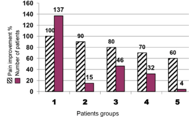
This study presenting the new acupuncture mandible microsystem, its anatomical representation on the face and its role in treating different acute painful disorders in the body. It is the latest microsystem discovered after about eighteen known microsystems throughout the history of acupuncture. It is a mini anatomical copy of the human body with unique distribution of body parts, the brain, the Spinal cord and the autonomic nervous system on the bilateral sides of the face. It differs from other microsystems on which body parts are not arranged in a systematic way like the human body anatomy. All the patients received acupuncture treatment. The main group of 234 patients received treatment in points on the mandible microsystem and the control group of 35 patients received treatment in a non-acupuncture point near the mandible microsystem. All The main group patients showed significant pain relief after the first treatment sessions, with varying degrees between 50 to 100 %, Compared with the sham effect which was 44% of the analgesic response. Numeric Rating Scale (NRS) for pain improved between 0 (no pain) and 3 (mild pain), which was not irritating to the patients and hence they returned to work and their normal daily life. Of the main group 137 patients showed 100% pain relief. 15 showed 90%, 46 showed 80%, 32 showed 70% and 4 showed 60% improvement. Follow-up showed improvement of functional and quality of life with no adverse effects or relapse. For the first time, we can use a technique for relieving pain in most parts of the body, with reproducible effects. Mandible microsystem is a unique in its anatomy and a new method of treating pain.
Introduction
Acupuncture Microsystems are regions of the body that contain maps of the entire organism with specific points or areas that correspond to each part. Ear acupuncture is the best-known system. Stimulating these points and areas has a rapid and powerful regulatory effect on energy and blood, they improve the function of the corresponding part of the body and they can be successfully used to relief any acute pain [1]. Microsystems are considered holographic images of the entire individual projected onto a particular part of the body. The ear, hand, scalp, foot, face, tongue, and the iris are some of the commonly recognized acupuncture microsystems [2]. Before the discovery of the holographic theory, man had already found that in specific areas of the body, diseases in any part of the body could be diagnosed and treated, such as ear, face, iris, nose and foot. Each part contains all the patho-physiological information of various regions of the whole body [3]. According to holographic theory, any abnormal changes have an effect both on a specific organ and on the holographic units in the other parts of the body. These holographic units can be used to diagnose and treat diseases [4]. The number of newly discovered microsystems is unlimited [5]. In 1973, Zhang developed the bio holographic theory. Dale was the first who suggested that, not only the ear, but every part of the body can function as a complete system for diagnosis and therapy [6].
In 1976, he introduced fourteen microsystems in the ear, hand, foot, abdomen, back, neck, scalp, face, nose, wrist, ankle, arm, leg and eye [7]. Since Dale the scientists developed these microsystems and discovered new ones. Up till recent time there are about eighteen acupuncture microsystems have been discovered in the world [4]. This study present the anatomy of the mandible microsystem and its relations to the mandible and the structures of the face. It also proved its role in relief of different acute painful conditions.
Why we need the mandible microsystem
Ear microsystem had about 8 classifications for distribution of body parts. For example liver point had 8 different sites on the ear, one for liver and the other 7 sites on the sites of other organs and also every other organ had 8 different sites [8-15]. This disturbed practitioners and practice itself. That is why since about 20 years I am thinking in a microsystem that contain all body parts in one fixed place not liable to change by any scientist.
Anatomy of the mandible
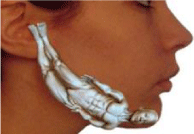
The mandible is the lower jaw and contain the lower teeth. It consists of two symmetrical halves. The mandible halves were separated at birth and their fusion completed by the end of first year. Each half is composed of horizontal part called the body and a vertical part called the ramus. The anterior triangular part of the body of the mandible is the mental process which forms the bony chin. The connection of the body and the ramus is the angle of the mandible. The upper end of the ramus extends to form the coronoid process anteriorly and the condyloid process posteriorly. The notch of the mandible separates the condyloid process from the coronoid process. Each half of the mandible is extending from the symphysis up to the condyloid and coronoid processes. The symphysis is the fusion line of the two halves of the mandible. Each half is called a hemi mandible and is separated from the other half at birth, so it can be considered as a micro system that carries all the information of the body like ear, hand and foot (Figure 1) [16,17].
Materials and Methods
This study included 234 patients, 119 males and 115 females. Their ages were between 14 – 68 years with mean age of 47 years. After committee acceptance, we started this prospective study on 2 groups: a main group of 234 patients and a control group of 30 patients. Each patient had acute pain in an area of body and received acupuncture treatment in points or areas on the mandible corresponding to anatomical pain regions in the main group and in the control group in a non-acupuncture point, one finger breadth below the small intestine 18 point (Quanliao) which lie directly below the outer canthus of the eye on the lower border of the zygomatic bone.
All patients had acute pain (less than 3 months): In the main group 45 patients had pain in the neck, 38 in shoulders (14 bilateral, 12 right, 12 left), 22 in upper limbs (5 arm, 9 elbow, 3 wrist, 3 hand, 2 fingers), 44 in trunk and 85 in lower limbs (31 sciatica, 29 knee, 8 leg, 6 ankle, 5 heel 2 foot, 2 sole, 2 toes). Numeric Rating scale (NRS) for pain [18] was evaluated before and after the first session. Successful treatment was evaluated by no recurrence of pain, functional improvement and quality of life. Monthly follow up visits were made for 3 months and the condition of each patient was reevaluated to document any relapse or adverse effects.
Mandible is a newly discovered microsystem
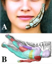
The mandible, used as a micro acupuncture system which has the same physiological functions that can regulate the human body functions through stimulation of their micro organs and structures (Figure 2). The treatment through mandible microsystem can be proved easily in pain therapy by any trained practitioner. It was successful with me for nine years of daily clinical practice. Because of absence of any facilities I had no choice to prove the anatomical representation of this system except by the clinical testing of the sites of the organs and areas of the body. It was the most suitable method to prove the effectiveness of this new system and its high accuracy rate. So, if the patient suffered of pain, an acupuncture needle is inserted in the skin of the corresponding part on either half of the mandible, Pain improved directly with varying degrees. This means that the site of the corresponding point or area is correct. I tested nearly every part of the body during these nine years mainly for pain and also for other diseases and symptoms.
Mandible is a remote-control system
This microsystem is similar to original human body but in mini parts which control the functions of the original macro parts and repair them, if disturbed, by four systems; (i) two separate complete control systems on either halves of the mandible, (ii) two central nervous systems including brain and spinal cord, (iii) two microsystems of mini parts that regulate the functions of the macro original parts of the body and (iv) two autonomic nervous systems which control the functions of most organs of the body. No other microsystem discovered up till now, can explain all the anatomical and physiological functions of the body like this system because it is a mini copy of the human body.
Therapy technique
The needle is inserted in the skin, connective tissue and muscles over the mandible. The skin of the face varies in thickness. It is thin around the eyes, thick over the forehead, temples and around the mouth and thickest over the chin. The muscles of the face are embedded within the connective tissue under the skin [19]. The needle is stimulated for one minute or more by even method that means moving the acupuncture needle to and fro and up and down movements at the same time. A 32 or 34-gauge filiform needle, 2.5 or 4 cm in length is used. The skin of the mandible is thin in comparison to other areas like the abdomen or thigh, so only the horizontal and oblique techniques are used. Needles are retained for 10 to 30 minutes after the energy arrival to the point.
Anatomical landmarks of mandible microsystem
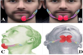
There are six vertical lines that determine the main anatomical areas of the body (Figure 3). 1. Vertical line in the anterior midline of the body from midpoint of lower lip to the lower border of the mandible, 2. Vertical line from medial angle of the eye to the lower border of the mandible, 3. Vertical line from lateral angle of mouth to the lower border of the mandible, 4. Vertical line from the center of the pupil of the eye to the lower border of mandible. 5. Vertical line from the lateral angle of the eye to the lower border of mandible and 6. Vertical line from the lateral end of the eye brow to lower border of mandible.
Body representation on the mandible
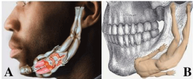
Head and Face: The head with the face are represented on the mental process. Its medial border is the midline of the body. Its lateral border is the vertical line that extend from the medial angle of the eye to the lower border of the mandible. Its upper border is the transverse crease of the chin and its lower border is the lower border of the mandible (Figure 4).
Head and Face: The head with the face are represented on the mental process. Its medial border is the midline of the body. Its lateral border is the vertical line that extend from the medial angle of the eye to the lower border of the mandible. Its upper border is the transverse crease of the chin and its lower border is the lower border of the mandible (Figure 4).
Secondly, the Spinal Cord - The spinal column and spinal cord inside lie on the lower border of the mandible. It extends from the level of the lateral angle of the mouth to the lateral angle of the mandible. It is divided into anterior half which includes the cervical and thoracic vertebrae and segments and posterior half which includes the lumbar, sacral and coccygeal vertebrae and segments. The spinal cord fills the spinal canal as in the three months old fetus in which the spinal cord occupies the whole length of the spinal canal [20-22], (Figure 6).
Thirdly, the Autonomic nervous system - The autonomic nervous system is represented in the brain stem and on both sides of spinal cord area on the lower border of the mandible similar to the anatomy of the human spinal cord and brain stem.
Fourthly, the Neck - The neck occupies an area of half an index finger breadth on both sides of the cervical Vertebrae on the lower border of the mandible from the level of the medial angle of the eye to the level of the angle of the mouth.
Fifthly, the Trunk - The trunk is divided into the chest, abdomen and pelvis. It is represented on both sides of the border of the mandible for an index finger breadth. From the level from lateral angle of mouth to the angle of the mandible.
Sixthly, the Upper Limb - The arms extend for half an index finger breadth on both sides of the chest, abdomen and pelvis areas. The shoulder lies at the level of the lateral angle of the mouth. The elbow lie at the level of the lower border of the chest midway between wrist and shoulder. The wrist is at the lower end of abdomen at the level of the lateral angle of the eye. The hand is between the lower end of the abdomen and the lateral angle of the mandible.
Seventhly, the Lower Limb - The two legs lie along the right and left halves of the ramus of the mandible. Each along its whole length from the angle of the mandible to the level of the helix crus of ear (Figure 7). The hip joint is at the level of the lateral angle of the mandible. The knee joint is at the midpoint between the hip and ankle joints. The ankle joint is at level of the lower border of the inter-tragic notch of the ear. The feet lie anterior to the ear. Each foot occupies an area of one index finger breadth from the level of the lower border of the cartilage of the inter-tragic notch to the level of the transverse helix crus.
Techniques of relieving pain in the mandible microsystem: For the first time in the history of acupuncture we can use a method for relieving pain, in most parts of the body, through three centers) local, spinal and brain centers) on each half of the mandible which are not known in any microsystem. The following is an example of treating left shoulder pain using these 6 centers (Figures 8-10).
Results
This study included 234 patients, 119 males and 115 females. Their ages were between 14 -68 years with mean age of 47 years. In the main group the mean NRS before and after the first session was 7.90 and 1.00 with pain relief percent of 87.17. The minimum and maximum NRS was 7 and 10 before the first session and 0 and 4 after it with pain relief percent between 50 and 100. In The control group the mean NRS before and after the first session was 8.90 and 7.33 with pain relief percent of 17.03. The minimum and maximum NRS was 8 and 10 before the first session and 5 and 8 after it with pain relief percent between 0 and 44. In the main group the significant improvement in function and in pain relief was between 50 to 100 %, compared with sham effect which was 44%.
Relation between session numbers and pain duration
There was a significant increase in session numbers with increase of pain duration (Figure 11).Relation between patient’s groups improved after the first session and their numbers with their improvement percentage
All the 234 patients showed pain relief after the first session with varying degrees. 137 patients showed complete relief of pain (100%), 15 showed 90% improvement, 46 showed 80% improvement, 32 showed 70% improvement and 4 showed 60% improvement (Figure 12).
Relation between number of sessions and number of patients with their percent
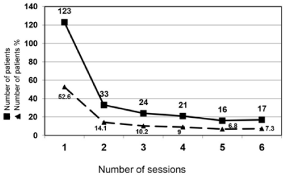
123 patients improved after one session, 33 improved after two sessions, 24 improved after three sessions, 21 improved after four sessions, 16 improved after 5 sessions and17 after 6 sessions (Figure13).
Number of patients treated through local area, spinal cord area, brain areas or combinations of them
Pain relieved in 35 patients using local areas only, in 77 patients using spinal cord centers, in 44 patients using brain motor and sensory areas and in 78 patients using either 2, 3, 4, 5 or 6 combinations of local, spinal cord or brain centers or areas (Table 1).
Discussion
Mandible microsystem is a micro human anatomy system on which the structures of the body are represented in the same positions as in the human body anatomy. The mandible, as a micro anatomy system, can be used as a micro acupuncture system which has the same physiological functions that can regulate the human body functions through stimulation of their micro organs and structures. This system is unique and new because it treats pain along its pathway by three ways:
1. Stimulating the primary site of pain in the body.
2. Stimulating its representing area on the spinal cord.
3. Stimulating its controlling motor and sensory areas in the brain.
Stimulation of any area of the body can affect the superficial and deep structures in this area from the skin on the front of the body to the skin on the back of the same area and correct any functional disorders in them. This system differs from all other microsystems in that the brain and spinal cord are represented anatomically in the same way as in modern medicine. Also, the right and left halves of the brain are represented on both halves of the mandible, so double stimulation of the brain can improve the results of therapy. Also, double representation of the body on bilateral halves of the mandible means that we can use two methods of treatment at the same time which increases the chances of cure of most pains and diseases. Throughout the nine years of practice, using this method the results were clear cut immediate or within minutes which make this system of value to be added to other microsystems.
Conclusion
Mandible microsystem is a functional image of the whole body in well-defined localized areas. Representation of the body parts on the mandible is correct and obeys the rules of the normal anatomy of the body. Furthermore, treatment of acute pain in any body area had reproducible effects.
Acknowledgments
The authors wish to thank all patients for giving us their written consent for inserting the acupuncture needles in their skin and took photos for needles positions.
- Starwynn D. Rapid Cancer Pain Relief with Auricular Micro-Macro technique. Acupuncture Today. 2008; 09. https://goo.gl/jC3u6T
- Soliman NE, Frank, BL Soliman Frank 3 Phase hand acupuncture. A Journal for Physicians by Physicians. 2005; 17: 29-34.
- Zhang Y. ECIWO Biology and Medicine: A New Theory of Conquering Cancer and a Completely New Acupuncture Therapy. China: Neimenggu People’s Press; 1987; 217. https://goo.gl/3F1sxj
- Wang Y. Micro- Acupuncture in practice. Churchill Livingstone. Elsevier. 2009; 274. https://goo.gl/d2jRNq
- Bouevitch, V. Microacupuncture Systems as Fractals of the Human Body. Medical Acupuncture. A Journal for Physicians by Physicians. 2003; 14: 14-16.
- Oleson, T. Auriculotherapy Manual Chinese and Western Systems of Ear Acupuncture. 3rd Ed. 2003. Reprent. 2009.
- Dale R. The Micro Acupuncture Systems, Part I & II. Am J Acupuncture. 1976: 4: 196- 224.
- Oleson, T. Auriculotherapy Manual, Chinese and Western Systems of Ear Acupuncture. 3rd Ed. 2003. https://goo.gl/8LS8jW
- Akerele D. WHO and the Development of Acupuncture Nomenclature, Overcoming a Tower of Babel. Am J Chinese Med. 1991; 1: 89-94. https://goo.gl/72iLoR
- WHO: A Standard International Acupuncture Nomenclature: Memorandum from a WHO meeting. WHO Bulletin. 1990; 68: 165-169. https://goo.gl/CZHpd3
- Wexo M. The Ear Gateway to Balancing the Body: A Modern Guide to Ear Acupuncture. New York: ASI Publishers; 1975. https://goo.gl/yEQtNm
- Essentials of Chinese Acupuncture: Ear Acupuncture Therapy. 1st Ed. 1980. 399-415. https://goo.gl/HwucbK
- Chen K, Cui Y. Handbook to Chinese Auricular Therapy. 1st Ed. Beijing, China: Foreign Languages Press; 1991. https://goo.gl/cqCGQf
- Acupuncture, A comprehensive Text: Ear Acupuncture. 5th Ed. pp 472-476, 1987. https://goo.gl/MKJdYT
- Rubach A. Principles of Ear Acupuncture. Microsystem of the Auricle. 2001. https://goo.gl/uyvHg3
- Hugo L O, Hans U . Mandibular Growth Anomalies. 2001. 17-18. https://goo.gl/jrrkA6
- Lewis A. Gray's Anatomy of the Human Body: The Mandible (Lower Jaw). 20th Ed. II. Osteology. 5b. 8. The Mandible (Lower Jaw). 1918. https://goo.gl/ko4tsB
- Childs J D, Piva S R, Fritz J M. Responsiveness of the Numeric Pain Rating Scale in Patients with Low Back Pain. Spine. 2005; 30: 1331-1334. https://goo.gl/T1j7rd
- Clemente, D. Clemente′s Anatomy Dissector. 2nd Ed. Carmine. 2007; 304. https://goo.gl/THkG3M
- Charles R, et al. The Human Nervous System: Structure and Function. 6th Ed. Springer. 2007; 12. https://goo.gl/ymqhWe
- Cochard R. Netter's Atlas of Human Embryology. The Nervous System. Elsevier Health Science. 2012. 63. https://goo.gl/ceysoG
- Vernon L. Spinal Cord Medicine: Principles & Practice. Demos Medical Publishing, 2nd Ed. 2010. 4. https://goo.gl/SHoHeF






Sign up for Article Alerts