Ectopia Lentis in a 2- Year - Old Child with Marfan syndrome?
Hannah Muniz Castro1*, Audrey Xi Tai1,2 amd Marjan Farid1,2
1University of California, Irvine School of Medicine, Irvine, California, USA
2Gavin Herbert Eye Institute, Irvine, California, USA
*Address for Correspondence: Hannah Muniz Castro, 3 Exeter, Irvine CA 92612; Tel: 760- 464-4773; E-mail: [email protected]
Submitted: 05 May 2017; Approved: 24 May 2017; Published: 29 May 2017
Citation this article: Castro HM, Tai AX, Farid M. Ectopia Lentis in a 2- Year - Old Child with Marfan syndrome. Int J Ophthal Vision Res. 2017;1(1): 012-015.
Copyright: © 2017 Castro HM, et al. This is an open access article distributed under the Creative Commons Attribution License, which permits unrestricted use, distribution, and reproduction in any medium, provided the original work is properly cited
Download Fulltext PDF
Purpose: We report a rare case of a 2 - year-old child with ectopia lentis and potential Marfan syndrome (MFS) and discuss her management.
Methods: A retrospective audit of a random sample of 500 patients with known proliferative diabetic retinopathy (PDR or R3), and 500 non-proliferative diabetic retinopathy (NPDR or R2). Images were re-assessed according to the English program criteria for DR levels using 1-FOV.
Results: On presentation, the patient did not exhibit any remarkable marfanoid skeletal features. The patient underwent a full ophthalmologic examination. Slit lamp examination revealed deep anterior chambers, superotemporal lens subluxation, and stretched and intact zonules in both eyes (OU). The patient was referred to genetics for gene testing and found to have a c.364C > T mutation in the FBN1 gene, supporting a diagnosis of Marfan syndrome. Due to a high irregular lenticular astigmatism, inability to obtain a functional manifest refraction for glasses, and high risk of refractive amblyopia, she was referred to us for lensectomy and secondary intraocular lens placement.
Conclusion: Ectopia lentis is the most common ophthalmologic finding of MFS, but does not commonly present in pediatric patients this young. It is important to investigate genetic and metabolic causes in order to accurately diagnose and determine appropriate medical management for each patient. In cases of amblyogenic lens dislocations, an early lensectomy and secondary scleral fixated intraocular lens placement is a feasible option.
Introduction
Ectopia lentis, or lens subluxation / dislocation, is a rare condition but is one of the most common ophthalmologic manifestations of Marfan Syndrome (MFS). Because laxity of the zonules takes time to develop, ectopia lentis is rarely diagnosed in children. We present a case of a 2 - year old with ectopia lentis and potential MFS and describe her management.
Case Description
A 2 - year old female was brought in by her mother after noticing that she watched television up close and held objects near her eyes. She also noticed increased blinking and preferred use of her left eye. The patient had no history of pain or trauma, no past medical history, and took no medications. She had no previous ophthalmologic exam and was further referred to the University for higher level care and management of bilateral ectopia lentis. The child’s family history was negative for ectopia lentis and MFS.
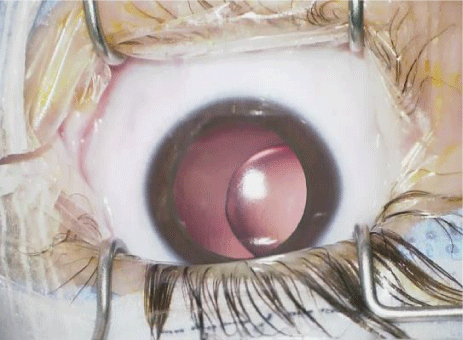
On presentation, the patient appeared her stated age, with normal weight and height. She did not exhibit any remarkable marfanoid skeletal findings. On exam, the patient’s vision was fix, follow, and maintain. Teller cards revealed binocular visual acuity of 20 / 130. Both eyes (OU) were soft by digital palpation. Dilated exam revealed deep anterior chambers OU. Lenses were displaced superotemporally OU (Figure 1). The zonules were stretched but remained intact. Cycloplegic retinoscopy exam revealed highly myopic refraction of - 12.50 + 2.00 x 175 of the right eye and - 12.50 + 2.00 x 5 of the left eye with irregular and unreliable astigmatism related to ectopic lenses. Fundus exam was normal. The patient was referred for genetic testing and prescribed temporary glasses pending further workup.
Genetic testing revealed a c.364C > T mutation in the Fibrillin - 1 (FBN1) gene, supporting a diagnosis of potential MFS characterized by predominant ectopia lentis with mild marfanoid phenotypes. The sequence variant was present in the heterozygous state and is associated with MFS inherited in an autosomal dominant pattern. Cardiology evaluation including EKG and echocardiogram were performed and found to be normal. The patient met criteria for “potential MFS” under the revised Ghent nosology [1] given an identified FBN1 mutation without aortic root dilation.
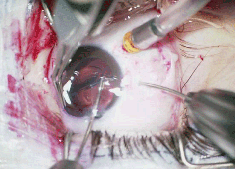
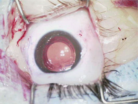
After discussion of the risks, benefits, and adverse effects of surgery, the patient’s parents desired surgical management for her ectopia lentis to prevent amblyopia. The patient underwent pars plana vitrectomy, lensectomy, and secondary Intraocular Lens (IOL) placement with scleral glue tunnel fixation in the left eye. Intraoperatively, the lens - capsule complex was removed in its entirety including the capsule and zonules through the pars plana approach. The iris diaphragm was noted to be very mobile. This is possibly a manifestation of the patient’s marfanoid syndrome. A posterior vitreous detachment was induced to separate the vitreous base from the macula and posterior pole, but the remaining vitreous was left in place to prevent retinal tears. Close shaving of the vitreous base was avoided to prevent complications. In preparation for the scleral glue tunnel fixation technique, as previously described by Dr. Amar Agarwal [2], conjunctival peritomies were made at 3 and 9 o’clock. Partial thickness scleral flaps were created and a 27 - gauge needle was used to create scleral tunnels where the haptics would be buried. A 23 - gauge needle was then used to create a full thickness sclerotomy under the flap and about 2 mm posterior to limbus. A 3- piece acrylic IOL was then injected into the anterior chamber through a standard 2.75 mm clear corneal incision. The leading haptic was grasped with microincision forceps through the sclerotomy, pulled out, and fed into the scleral tunnel. Using the handshake technique, the trailing haptic was placed into the eye and pulled through the second sclerotomy in the same fashion and placed into its tunnel (Figure 2). The haptics were secondarily secured to the sclera with 8 - 0 vicryl sutures, which also helped to close the sclerotomy. Tisseel glue was used to secure down the scleral flaps as well as the overlying conjunctival flaps. A 10 - 0 vicryl suture was used to close the corneal incision. The intraocular lens was noted to be well centered and secure at the end of the case (Figure 3). One month later, the patient underwent the same operation for the right eye without any complications. At the last evaluation 4 months after the last operation, visual acuity was 20 / 60 OU with glasses. The patient was doing well and her parents had no other complaints.
Discussion
This case represents one of the few rare cases of a young child with potential MFS presenting with dislocated lens and amblyogenic risk from high lenticular astigmatism. Excess laxity of the zonule fibers is a gradual process and therefore severe visually significant dislocation of the lens is rarely diagnosed in children [3]. Few cases of young children with ectopia lentis associated with FBN1 mutations have been reported. Zadeh et al [4], reported 8 cases of ectopia lentis presenting as the primary feature in MFS. The youngest patient was diagnosed with bilateral ectopia lentis at age of 2 - years which was surgically managed at age 3 - years. She had mild marfanoid skeletal deformities as well as an echocardiogram demonstrating aortic root dilation at the upper limits of normal. Zhang et al [5], reported a case of early onset ectopia lentis due to an FBN1 mutation with non - penetrance in a 6 - year - old boy. In a review of 74 cases with FBN1 mutations, Arbustini et al [6], reported on two children less than 2 - years old with ectopia lentis.
FBN1 is found on chromosome 15q21.1 and encodes the gene for fibrillin, a large glycoprotein that forms micro fibrils found in multiple tissues. A wide variety of mutations result in Marfan syndrome and other related fibrillinopathies that exhibit autosomal dominant inheritance. Lens dislocation is thought to be caused by structural deficiencies in fibrillin, resulting in disrupted microfibrillar assembly and defective zonular fiber production [7]. Although most individuals with MFS have a positive family history, about 25 % of cases arise from de - novo mutations, as in this patient we present [8]. The c.364C > T missense mutation found in our patient substitutes codon 122 coding for cysteine with one coding for arginine, a mutation that has been previously described and is associated with MFS with predominant ectopia lentis [9].
Management of ectopia lentis in children focuses on optimizing visual acuity to prevent amblyopia. Surgery is indicated when spectacle correction does not provide the child with adequate visual acuity for daily functioning or if there are risks of development of amblyopia without surgical intervention. In this case, the decision to undergo operative management was made due to decreased distance visual acuity of both eyes with spectacle correction, high myopia, and prevention of amblyopia. Surgical options for ectopia lentis include intracapsular cataract extraction, lensectomy with pars plana vitrectomy, and phacoemulsification with a Cionni - modified capsular Tension Ring (CTR) [10]. Visual rehabilitation after removal of the dislocated lens can be next achieved with IOL placement or aphakic with spectacle correction. Surgical management in MFS involves additional challenges and the most dreaded complication is Retinal Detachment (RD). In a review of 64 eyes in 39 patients with MFS who underwent surgery, severity of lens dislocation and higher axial myopia were statistically significant risk factors for the development of RD postoperatively [10]. The predisposing pathologic factors leading to the development of RD in MFS include an abnormally distended sclera, increased axial length, zonular insufficiency with ectopia lentis, abnormal vitreous equatorial attachments, and liquefied and mobile vitreous in an enlarged eye [11]. We present a rare case of spontaneous MFS in a 2 - year old that required lensectomy and secondary IOL placement. Due to the high risk of refractive amblyopia in the pediatric population, early lensectomy and secondary scleral fixated IOL placement is a suitable option.
Acknowledgements
The authors have no relevant disclosures, proprietary interest, or conflict of interest related to this study. This research received no grant or financial support from any public, commercial, or nonprofit funding agencies.
- Loeys BL, Dietz HC, Braverman AC, Callewaert BL, De Backer J, Devereux RB, et al. The revised Ghent nosology for the Marfan syndrome. J Med Genet. 2010; 47: 476-485. https://goo.gl/JQdwIR
- Agarwal A, Kumar DA, Jacob S, Baid C, Agarwal A, Srinivasan S. Fibrin glue-assisted sutureless posterior chamber intraocular lens implantation in eyes with deficient posterior capsules. J Cataract Refract Surg. 2008; 34: 1433-1438. https://goo.gl/xtRGnn
- Dureau P. Pathophysiology of zonular diseases. Curr Opin Ophthalmol. 2008; 19: 27-30. https://goo.gl/Z3RwCS
- Zadeh N, Bernstein JA, Niemi AK, Dugan S, Kwan A, Liang D, et al. Ectopia lentis as the presenting and primary feature in Marfan syndrome. Am J Med Genet A. 2011; 155A: 2661-2668. https://goo.gl/TAH7L8
- Zhang L, Lai YH, Capasso JE, Han S, Levin AV. Early onset ectopia lentis due to a FBN1 mutation with non-penetrance. Am J Med Genet A. 2015; 167:1365-1368. https://goo.gl/rILmCb
- Arbustini E, Grasso M, Ansaldi S, Malattia C, Pilotto A, Porcu E, et al. Identification of sixty-two novel and twelve known FBN1 mutations in eighty-one unrelated probands with Marfan syndrome and other fibrillinopathies. Hum Mutat. 2005; 26: 494. https://goo.gl/X1OaS2
- Nemet AY, Assia EI, Apple DJ, Barequet IS. Current concepts of ocular manifestations in Marfan syndrome. Surv Ophthalmol. 2006; 51: 561-575. https://goo.gl/L60UDS
- Judge DP, Dietz HC. Marfan's syndrome. Lancet. 2005; 366:1965-1976. https://goo.gl/XAZgxw
- Jin C, Yao K, Sun Z, Wu R. Correlation of the recurrent FBN1 mutation (c.364C > T) with a unique phenotype in a Chinese patient with Marfan syndrome. Jpn J Ophthalmol. 2008; 52: 497-499. https://goo.gl/jOCTcF
- Fan F, Luo Y, Liu X, Lu Y, Zheng T. Risk factors for postoperative complications in lensectomy-vitrectomy with or without intraocular lens placement in ectopia lentis associated with Marfan syndrome. Br J Ophthalmol. 2014; 98:1338-1342. https://goo.gl/iDDMjC
- Dotrelova D, Karel I, Clupkova E. Retinal detachment in Marfan's syndrome. Characteristics and surgical results. Retina. 1997; 17: 390-396. https://goo.gl/XJi3kQ


Sign up for Article Alerts