Orbital Cyst with Ependymal Differentiation Associated with Microphthalmia?
Maria D. Garcia*, Diva R. Salomao and Lilly H. Wagner
Department of Ophthalmology, Mayo Clinic, 200 1st ST SW, Rochester, MN 55905, United States
*Address for Correspondence: Maria D. Garcia, Department of Ophthalmology, Mayo Clinic, 200 1st ST SW, Rochester, MN 55905, United States, Tel: +507-284-2233; E-mail: [email protected]
Submitted: 26 November 2019; Approved: 16 December 2019; Published: 19 December 2019
Citation this article: Garcia MD, Salomao DR, Wagner LH. Orbital Cyst with Ependymal Differentiation Associated with Microphthalmia. Int J Ophthal Vision Res. 2019;3(1): 013-015.
Copyright: © 2019 Garcia MD, et al. This is an open access article distributed under the Creative Commons Attribution License, which permits unrestricted use, distribution, and reproduction in any medium, provided the original work is properly cited
Download Fulltext PDF
Introduction
We report the case of a male infant who presented with an orbital cyst associated with microphthalmia. On exam, there was a large cystic lesion in the right orbit with no visualization of the globe. Magnetic resonance imaging demonstrated a small globe, which was displaced posteriorly by the orbital cyst. The cyst was excised and the histology revealed it was lined focally by ependymal cells, which indicates formation during an early stage of embryologic development. Orbital cysts associated with microphthalmia are colobomatous lesions that typically present unilaterally with no associated systemic findings. We report a case of a 5-month-old infant with such findings.
Case Report
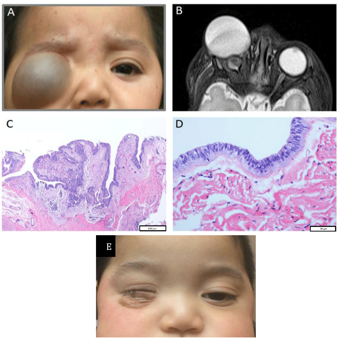
A full-term male infant, born at an outside institution, was found to have a large mass in the right inferior orbit at birth. At 5 months of age, the patient presented with his parents to our oculoplastic surgery service at [redacted for review purposes] for further evaluation (Figure 1A). At the outside facility, he was noted to have preauricular pits, but no other systemic anomalies. There was no family history of ocular disorders. On the side of the orbital cyst, the globe could not be visualized. Fundus examination of the contralateral eye showed an inferior chorioretinal coloboma sparing the macula. Genetic testing was performed for possible CHARGE syndrome and he was found to have a copy number gain of unknown clinical significance within chromosome 3q29. Serum titers for cytomegalovirus and toxoplasmosis were negative. At our institution, we performed magnetic resonance imaging. This revealed a well-defined cystic lesion in the anterior right orbit adjacent to a small globe, which was displaced posteriorly (Figure 1B).
After discussing the poor visual potential for the malformed eye and possible communication with the cyst which may necessitate enucleation, the parents wished to proceed with excision of the cyst. A right orbitotomy with removal of the cyst and reconstruction with a free periumbilical fat graft were performed. A conformer was placed and secured with a temporary tarsorrhaphy. The globe did not communicate with the cyst, and was therefore left in place to stimulate growth of the bony orbit.
Histologically, the cyst wall consisted of fibrovascular tissue surrounding a unilocular cavity lined by disorganized neuroglial cells along with areas lined by cells with cubodial/cylindrical morphology suggestive of ependymal cells (Figure 1 C,D). The glial cells were positive for GFAP and weakly for S100 protein by immunostains. In the areas lined by a layer of cuboidal to cylindrical cells, the cells were positive for GFAP and Ker AE1/AE3, but negative for Olig2 and Cam 5.2. We hypothesize that the ependymal cells, which are usually lining the ventricles of the brain and spinal cord, are likely present due to formation of this cyst in early embryogenesis, around the same time the optical vesicles form as an outpouching from the primitive forebrain [1]. This would also explain the anterior location of the cyst compared to typical colobomatous cysts, which are posterior to the globe.
Two and a half months after surgery, the cyst had resolved and the previously distended lower eyelid skin was contracting (Figure 1 E). The patient is currently seeing an ocularist for evaluation of serial enlarging conformers in order to promote eyelid/fornix growth and allow fitting of a prosthesis around age three or four.
Discussion
Orbital cysts associated with microphthalmia are considered colobomatous lesions of unknown etiology [2]. A possible association with maternal vitamin A deficiency has been suggested [3,4]. Colobomatous orbital cysts usually present unilaterally at birth as an isolated finding [5,6]. Rare examples of bilateral cases have been reported and may be accompanied by systemic abnormalities involving the heart, CNS, lungs, kidneys or clefting defects [5,6]. The cyst wall is usually lined by disorganized neuroglial tissue. Although our patient had a chorioretinal coloboma in the contralateral eye, it is unclear if this is related to the orbital cyst as he did not exhibit any systemic abnormalities.
Different types of cystic orbital lesions can occur in children due to inflammatory, infectious or neoplastic processes. Shields and Shields have proposed a classification of orbital cysts of childhood [2]. The category of neural cysts includes orbital cysts associated with maldevelopment of the eye, as well as cysts associated with meningocele and meningo-encephaloceles [2]. Our case represents a distinct lesion that likely formed earlier during embryologic development than classic colobomatous cysts associated with microphthalmos.
Orbital cysts associated with maldevelopment of the eye can be managed expectantly, if the lesion is not causing discomfort or cosmetic deformity. Frequently, a scleral shell can be fitted over the microphthalmic globe, and the posterior cyst actually stimulates growth of the orbital bones. However, in our case the lesion appeared to cause pain, and its large size and anterior location would not have allowed for prosthesis fitting. After excision of the cystic lesion and reconstruction of the socket with an implant or autologous graft material, patients need to be followed closely by an ocularist for placement of serially enlarging conformers. The goal is to support growth of the eyelids and fornices to allow for fitting of a prosthesis by the time the child reaches school age.
- Fuhrmann S. Eye morphogenesis and patterning of the optic vesicle. Curr Top Dev Biol. 2010; 93: 61-84. PubMed: https://www.ncbi.nlm.nih.gov/pubmed/20959163
- Shields JA, Shields CL. Orbital cysts of childhood - classification, clinical features and management. Surv Ophthalmol. 2004; 49: 281-299. PubMed: https://www.ncbi.nlm.nih.gov/pubmed/15110666
- Hornby SJ, Ward SJ, Gilbert CE, Dandona L, Foster A, Jones RB. Environmental risk factors in the etiology of congenital malformations of the eye in children in South India. Ann Trop Paediatr. 2002; 22: 67-77. PubMed: https://www.ncbi.nlm.nih.gov/pubmed/11926054
- Decock CE, Breusegem CM, Van Aken EH, Leroy BP, Van Den Broecke CM, Delanghe JR. High beta-trace protein concentration in the fluid of an orbital cyst associated with bilateral colobomatous microphthalmos. Br J Ophthalmol. 2007; 91: 836. PubMed: https://www.ncbi.nlm.nih.gov/pubmed/17510479
- Foxman S, Cameron JD. The clinical implications of bilateral microphthalmos with cyst. Am J Opthalmol. 1984; 97: 632-638. PubMed: https://www.ncbi.nlm.nih.gov/pubmed/6720843
- Chaudhry IA, Arat YO, Shamsi FA, Boniuk M. Congenital microphthalmos with orbital cysts: distinct diagnostic features and management. Ophthalmic Plast Reconstr Surg. 2004; 20: 452-457. PubMed: https://www.ncbi.nlm.nih.gov/pubmed/15599246


Sign up for Article Alerts