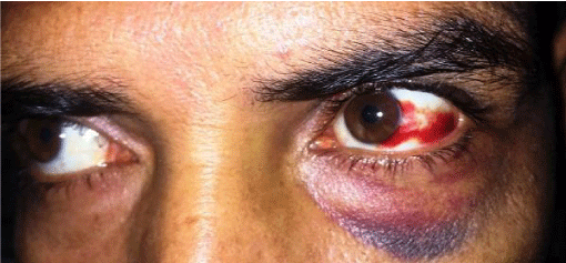
Traumatic Subconjunctival Haemorrhage?
Siddharth Madan*
Department of Ophthalmology, Lady Hardinge Medical College and Associated Hospitals, University of Delhi, New Delhi, India
*Address for Correspondence: Siddharth Madan, Department of Ophthalmology, Lady Hardinge Medical College and Associated Hospitals, University of Delhi, New Delhi, India, Tel: +91-971-121-2928; E-mail: [email protected]
Submitted: 02 December 2018; Approved: 12 December 2018; Published: 14 December 2018
Citation this article: Madan S. Traumatic Subconjunctival Haemorrhage. Int J Ophthal Vision Res. 2018;2(2): 033-034.
Copyright: © 2018 Madan S. This is an open access article distributed under the Creative Commons Attribution License, which permits unrestricted use, distribution, and reproduction in any medium, provided the original work is properly cited
Keywords: Subconjunctival haemorrhage; Trauma; Peri orbital ecchymosis
Download Fulltext PDF
Purpose: To evaluate the characteristics of color vision and relevant factors in normal myopic eyes and to assess changes in color vision before and after Laser in Situ Keratomileusis (LASIK).
Methods: Fifty-nine consecutive eyes of 31 patients were prospective valuated in a randomized contralateral-eye study. The color vision was measured with Farnsworth-Mun-sell 100-hue test (FM100 test), and relationships with some optic parameters were analyzed in normal myopic eyes. The difference between postoperative and preoperative was evaluated before and 1 day, 1 week and 1 month after surgery.
Results: For normal eyes, there was positive correlation between Square Root of Total Error Score (Sqrt TES) and intraocular pressures (p = 0.045). Sphere, astigmatism, and visual acuity preoperative showed no correlation with Sqrt TES. The Sqrt TES preoperative of the FM 100 test was 7.89 ± 2.34. After surgery, 1 day Sqrt TES was significant higher than the 1 month postoperative values (p = 0.018, ANOVA test). No significant change in the Sqrt TES was observed at 1 day (8.08 ± 2.47, p = 0.469, paired t test), 1 week (7.76 ± 2.16, p = 0.489, paired t test), or 1 month (7.61 ± 2.75, p = 0.146, paired t test).
Conclusion: Intraocular pressures have significant correlation with Sqrt TES in normal myopic eyes. Little changes were detected by the FM 100 test after Laser in Situ Keratomileusis (LASIK). The temporal changes of the photoreceptor layer of the retina maybe as a resulting of increase of intraocular pressures in LASIK surgery might explain this minimal influence.
Abbreviations
SCH: Subconjunctival Haemorrhage
Introduction
Ophthalmic problems are a common presentation in every step of daily clinical practice to general physicians and emergency services in hospitals. Red eye has many potential causes and every cause needs targeted intervention. Sudden onset Subconjunctival Haemorrhage (SCH) is a disturbing compliant for the patients who develop it. Causes for subconjunctival haemorrhage are multifactorial. Patients who are on anticoagulant therapy, have uncontrolled hypertension, diabetes, hematological dyscrasias or arteriosclerosis may develop SCH [1]. Trauma and contact lens usage are common causes in younger patients [2]. Causes for trauma may include a foreign body, a blunt injury or a penetrating injury to the globe or vigorous rubbing of the eyes. Penetrating eye injuries can cause SCH at all noticeable levels [3]. Fracture of the orbital bones may cause a delayed appearance of SCH that may be noticed after 12 hours upto as long as 24 hours. This delay in appearance of the haemorrhage results from leakage of the blood slowly under the conjunctival tissue [3]. Fracture of the base of skull needs to be ruled out in such clinical scenarios. This 35 year old patient presented with sudden onset infra-orbital ecchymosis and SCH localized temporally following a history of assault with a blunt trauma to the left eye (Figure 1). His visual acuity on presentation was 6/6 in both the eyes with normal ocular movements, pupils and fundus examination. No orbital or peri-orbital bony tenderness was elicited on clinical examination. The presentation in such cases is usually painless and is characterized by sharply defined area of redness in the subconjunctival space and the posterior extent of the haemorrhage is usually well demarcated to distinguish the same from retrobulbar haemorrhage. The traumatic SCH usually remains localized to the site of injury and is mostly temporal in location. Visual acuity remains normal in most cases unless it is associated with trauma to other structures of the anterior or posterior segment. The blood in the subconjunctival space resorbs spontaneously in 2 to 4 weeks. Most cases do not require treatment and only require reassurance to a disturbed patient. A recurrent SCH may need systemic work-up to localize underlying etiology however a good clinical history and comprehensive clinical examination averts the need for unnecessary investigations. This patient had a spontaneous recovery in 2 weeks and did not require further intervention.
Consent for photographs
Taken from the patient
- Frings A, Geerling G, Schargus M. Red Eye: A guide for non-specialists. Dtsch Arztebl Int. 2017; 114: 302-312. https://goo.gl/AbTrHd
- Tarlan B, Kiratli H. Subconjunctival hemorrhage: risk factors and potential indicators. Clin Ophthalmol. 2013; 7: 1163-1170. https://goo.gl/PQ7GGT
- Duke Elder S. System of ophthalmology diseases of the outer eye. VIII. London: Henry Kimpton; 1965. p. 34-39.


Sign up for Article Alerts