Hydrocephalus Induced Psychosis Associated with Tentorial Meningioma: A Case Report?
Enyinna L. Nwachuku1*, Ayodele Ogunmola3, Lawson Bernstein2, Matthew Pease1 and Daniel A. Wecht1
1Department of Neurological Surgery, University of Pittsburgh Medical Center, Pittsburgh, Pennsylvania
2Department of Psychiatry, University of Pittsburgh Medical Center-McKeesport Hospital, Pittsburgh, Pennsylvania
3University of Pittsburgh School of Medicine, Pittsburgh, Pennsylvania
*Address for Correspondence: Enyinna L. Nwachuku, Department of Neurological Surgery, UPMC Presbyterian-Suite B-400 200 Lothrop Street, Pittsburgh, PA 15213, Tel: 412-647-6777; E-mail: Nwachukuel@upmc.edu
Submitted: 30 December 2019; Approved: 10 January 2020; Published: 11 January 2020
Citation this article: Nwachuku EL, Ogunmola A, Bernstein L, Pease M, Wecht DA. Hydrocephalus Induced Psychosis Associated with Tentorial Meningioma: A Case Report. Sci J Neurol Neurosurg. 2020;6(1): 001-003.
Copyright: © 2020 Nwachuku EL, et al. This is an open access article distributed under the Creative Commons Attribution License, which permits unrestricted use, distribution, and reproduction in any medium, provided the original work is properly cited
Keywords: Meningioma; Posterior fossa; Tentorium cerebelli; Hydrocephalus; Psychosis
Download Fulltext PDF
Background: This report describes a unique case of a patient that developed psychotic symptoms believed to be secondary to a tentorial meningioma with associated hydrocephalus. These psychotic symptoms subsequently abated with placement of a ventriculoperitoneal shunt.
Case description: 60-year-old female was admitted to an inpatient psychiatric facility on a psychiatric involuntary commitment petition due to progressive paranoia, homicidal ideation and psychosis. The work up showed a calcified six cm tentorial meningioma with associated hydrocephalus. The patient initially rejected treatment but later became amenable to placement of Ventriculoperitoneal Shunt (VPS).
The patient underwent placement of right-sided parietal VPS, integra-medium pressure valve, without any complication. After the acute post-operative period, the patient was transferred back to the inpatient psychiatric service for management of her psychotic symptoms. During her course of care, the staff and family began to note a more rational thought process and her psychotic symptoms abated by time of discharge from the hospital which was within a few days after VPS placement. After resolution of her psychosis, patient’s tentorial meningioma was resected successfully after she gave informed consent.
Conclusion: Neuropsychiatric symptoms are rarely associated with posterior fossa meningiomas. The ventriculomegaly associated with the mass effect can be mitigated with Cerebrospinal Fluid (CSF) diversion in the form of ventriculoperitoneal shunt, which may also ameliorate the patient’s neuropsychiatric presentation.
Introduction
Tentorial meningiomas encompass 2-6% of all intracranial meningiomas [1-3]. These meningiomas are dichotomized into falcotentorial and torcular. Torcular meningioma are stated to be located in the posterior cranial fossa attached to the confluence of sinuses with or without sinus invasion. In contrast, falcotentorial meningiomas are described as being attached at the angle between the falx and the tentorium with or without invasion into the straight sinus. Many different surgical approaches are available as surgical technology advances and as more neurosurgeons are trained to safely and effectively resect these meningiomas. A majority of patients with these tentorial meningiomas present with headaches secondary to hydrocephalus and visual difficulties [3]. There is limited data in the literature to suggest that some patients can also develop psychiatric disturbance as a result of this pathology. This report describes a unique case of a patient that developed psychotic symptoms believed to be secondary to a tentorial meningioma with associated hydrocephalus. These psychotic symptoms subsequently abated with placement of a Ventriculoperitoneal Shunt (VPS).
Case Report
DM, a 60-year-old female with a past medical history significant for depression, anxiety, fibromyalgia, was admitted to an inpatient psychiatric facility on a psychiatric involuntary commitment petition due to progressive paranoia, homicidal ideation and psychosis. These included the delusional beliefs that her husband was unfaithful, had paramours living in the marital home, and was physically abusive to the patient and/or trying to poison her. As a result of this, police had been called numerous times to the patient’s home after she verbally abused female neighbors, calling them “whores” and claimed physical assault by the husband. The patient had also threatened to shoot her husband and/or these women as the result of her conjugal paranoid delusions.
During her initial intake evaluation, a Computed Tomography (CT) of the head obtained as part of her diagnostic evaluation revealed an approximately six cm calcified tentorial lesion, thought to be a meningioma with associated hydrocephalus. This meningioma was also noted two years prior during trauma evaluation after a motor vehicle accident, however it had increased in size with associated progressive hydrocephalus at the time of this admission. During the interval two years, the patient’s husband had noticed a steady decline in her overall mental status as delineated above.
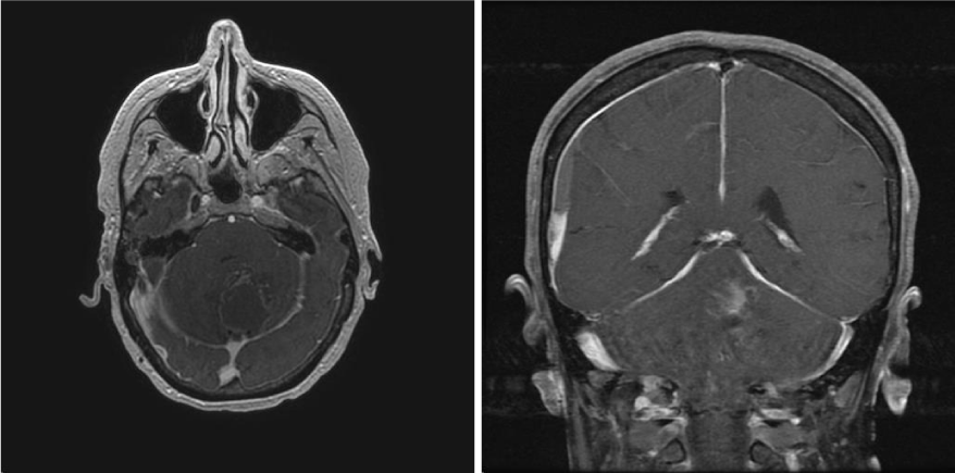
Given the progression of the meningioma with symptomatic hydrocephalus, neurosurgery was consulted for evaluation and management. A Magnetic Resonance Image (MRI) of the brain with and without gadolinium was obtain. It displayed the characteristic radiographic appearance of a meningioma i.e. homogeneous contrast enhancement, extra-axial, and associated dural tail, figure 2. The senior author (DAW) discussed extensively with the patient and husband the expanding posterior fossa meningioma as the etiology for her headaches and visual problems. Resection of the presumed meningioma was offered to the patient, which she vehemently refused. However, upon further discussion, the patient was agreeable to palliative cerebrospinal fluid diversion via ventriculoperitoneal shunt.
The patient underwent a right-sided parietal VPS with laparoscopic assistance without any complication, and an integra-medium pressure valve was placed. After the acute post-operative period, the patient was transferred back to the inpatient psychiatric service for management of her psychotic symptoms. During her course of care, the staff and family began to note a more rational thought process and her psychotic symptoms abated by time of discharge from the hospital which was within a few days after VPS placement. The dosage of pre-operative initiated antipsychotic medication Olanzapine was decreased from five mg qHs to two and a half mg qHs. With the noted improvement, the patient began to express interest in having her meningioma surgically removed.
The patient was seen for a two-week post-operative follow up for assessment and suture removal at which time the decision was made with the senior author to proceed with suboccipital craniectomy for tentorial meningioma resection. One month after her initially VPS placement, the patient was brought back to Presbyterian hospital of UPMC for the aforementioned surgery. A pre-operative head CT showed complete resolution of the patient’s hydrocephalus, figure 1. DM underwent a suboccipital craniectomy with a supracerebellar infratentorial approach to Grade III Simpson resection given that the superior aspect of the tumor was adherent to vital vascular structures such as the torcular and straight sinus, figure 3. Of note, in figure 3, there is a notable non-compressive right subacute subdural hematoma which was clinical observed and resulted in no clinical sequelae. Intraoperative pathology was consistent with Grade II atypical meningioma, Ki 67: < 1%. The patient’s hospital course was uncomplicated. She was able to be discharged to a rehabilitation facility on post-operative day three. Patient was at the rehabilitation facility for 8 days prior to discharge home. At the time of discharge, the patient was completely off her Olanzapine. The patient was seen in clinic at the two-week post-operative visits where she and the family endorsed no psychotic symptoms.
Discussion
The classic neurosurgical teaching regarding meningiomas for any pathologic grade is that 85% of meningiomas are grade me benign entities [4]. Typical patient presentation involves symptoms of headaches, seizures and transient paresis and paresthesia. Additionally, with associated hydrocephalus most patient have headaches, nausea, vomiting, blurry vision, but rarely pyschosis or delusional experiences. However, during the management of this patient, a significant amount of her presenting symptoms were neuropsychiatric in nature. This case report extends a small but growing literature that a minority of meningioma patients may present with primarily neuropsychiatric symptoms to include psychosis [4], as opposed to the classic symptoms delineated above. In this patient these neuropsychiatric symptoms, mainly delusional in nature, were abated in a progressive manner after the patient underwent a ventriculoperitoneal shunt. To our knowledge, there is very limited medical literature description of ventricular cerebrospinal fluid diversion ameliorating psychotic symptoms in a meningioma patient with obstructive hydrocephalus.
The pathophysiology for our patient’s psychosis is unknown, except to say that VPS prior to meningioma resection lead to rapid and complete psychotic symptom abatement. One might analogize that similar to normal pressure hydrocephalus ventriculomegaly associated cognitive dysfunction, our patient’s psychosis was simply the result of a global Central Nervous System (CNS) insult from her obstructive hydrocephalus and stretching of white matter tracks of the limbic system. However, this begs the question why this phenomenon does not commonly occur in other pathological processes that cause hydrocephalus.
Potential pathophysiologies for our patient’s presentation could include non-convulsive post-ictal psychosis [5], diaschisis related central nervous system dysfunction anatomically distant from the primary lesion and its associated effect [2] or psychosis associated with cerebellar disease process given our expanding understanding of its’ role in schizophrenaia. Additionally, slowly progressive brainstem compression secondary to hydrocephalus could lead to the altered level of mentation and/or perception of reality that is at time seen in patient with brainstem pathologies, such as central pontine myelinolysis. While the exact pathophysiology for our patient’s presentation ultimately remains unknown, this case demonstrates that psychosis associated with cerebellar meningioma related obstructive hydrocephalus can potentially be alleviated with cerebrospinal fluid diversion in the form of ventriculoperitoneal shunt.
Conclusion
Hydrocephalus is a known phenomenon associated with intracranial posterior fossa meningioma with significant mass effect. Neuropsychiatric symptoms are rarely associated with these meningiomas. The ventriculomegaly associated with the mass effect can be mitigated with CSF diversion in the form of ventriculoperitoneal shunt, which may also ameliorate the patient’s neuropsychiatric presentation.
- Aguiar PH, Tahara A, de Almeida AN, Kurisu K. Microsurgical treatment of tentorial meningiomas: Report of 30 patients. Surg Neurol. 2010; 1: 36. PubMed: https://www.ncbi.nlm.nih.gov/pubmed/20847917
- Bir SC, Sapkota S, Maiti TK, Konar S, Bollam P, Nanda A. Evaluation of ventriculoperitoneal shunt related complications in intracranial meningioma with hydrocephalus. J Neurol Surg. 2017; 68: 30-36. PubMed: https://www.ncbi.nlm.nih.gov/pubmed/28180040
- Bernard JA, Mittal VA. Cerebellar-motor dysfunction in schizophrenia and pyschosis-risk: The importance of regional cerebellar analysis approaches. Front Psychiatry. 2014; 5: 160. PubMed: https://www.ncbi.nlm.nih.gov/pubmed/25505424
- Hashemi M, Schick U, Hassler W, Hefti M. Tentorial meningioma with special aspect of the tentorial fold: Management, surgical technique, and outcome.Acta Neurochir. 2010; 152: 827-834. PubMed: https://www.ncbi.nlm.nih.gov/pubmed/20148271
- Ishii R, Canuet L, Iwase M, Kurimoto R, Ikezawa K, Robinson SE, et al: Right parietal activation during delusional state in episodic interictal psychosis of epilepsy: a report of two cases. Epilepsy Behav. 2006; 9: 367-372. PubMed: https://www.ncbi.nlm.nih.gov/pubmed/16884960


Sign up for Article Alerts