Two Cases of Hemorrhagic Stroke Following Snake Bite in Kara Teaching Hospital in a Semi Rural Area in Togo?
Kumako VK1*, N’Timon B2, Apetse K3, Guinhouya KM3, Agba L Th3, Assogba K3, Belo M3 and Balogou AK3
1Department of Neurology, University of Kara, Togo
2Department of Radiology, University of Kara, Togo
3Department of Neurology, University of Lome, Togo
*Address for Correspondence: Vinyo Kodzo Kumako, Department of Neurology, University of Kara, Togo, Tel: +002-289-012-3967; E-mail: vincent_kumako@yahoo.fr/vkumako23@gmail.com
Submitted: 01 March 2017; Approved: 16 April 2017; Published: 17 April 2017
Citation this article: Kumako VK, N’Timon B, Apetse K, Guinhouya KM, Agba L Th, et al. Two Cases of Hemorrhagic Stroke Following Snake Bite in Kara Teaching Hospital in a Semi Rural Area in Togo. Sci J Neurol Neurosurg. 2018;4(1): 001-004.
Copyright: © 2017 Kumako VK, et al. This is an open access article distributed under the Creative Commons Attribution License, which permits unrestricted use, distribution, and reproduction in any medium, provided the original work is properly cited
Download Fulltext PDF
Introduction: Envenomation is a public health problem in developing countries. Neurovascular complications are not exceptional.
Observations: We report two cases of hemorrhagic stroke which complicate an envenomation treated late.
The first patient was 27 years old woman, who had been admitted for right hemiparesis and aphasia two weeks after a viperidae bite. She was then treated with polyvalent antivenom (FAV-Afrique®).
The second patient was 31 years old man who was treated three days after the envenomation with a first dose of polyvalent antivenom (FAV-Afrique®). Twelve days later he presented left hemiparesis. He then received a second dose of polyvalent antivenom (FAV-Afrique®) because of supposed inefficiency of the first dose.
Both patients presented hemorrhagic stroke on CT-scan. They received symptomatic treatment with re-education. The outcome was favorable for both.
Conclusion: The morbidity and mortality related to envenomation by snake bite remains high in our environments. The neurovascular complications of these bites are often severe. Prevention by raising the awareness of target populations and better organization of our health systems will reduce the incidence and complications of these envenomation
Introduction
Snake bite envenoming is a major public health problem in the sub-Saharan African. Morbidity and mortality are high because of the difficulties of access to care, especially antivenom (FAV-Afrique®), poverty and ignorance [1]. In sub-Saharan Africa, about 314 000 bites are recorded annually with about 7 300 deaths [2,3].
Envenomation by viperids causes visceral complications that are accompanied by sequelae in 1 to 10% of cases [4]. Serious neurological complications, such as muscle paralysis and stroke, are related to the toxic effects of venom on neuromuscular transmission and hemostasis [5].
We report two clinical cases of hemorrhagic stroke following a viperid bite recorded at Kara Teaching Hospital.
Case 1
A 27-year-old woman with unknown cardiovascular risk factor was referred to, Kara Teaching Hospital Neurology Unit from a Peripheral Care Unit for sudden right hemiplegia with aphasia evolving 24 hours before admission.
The patient had been bitten on her left foot by a viperid two weeks before. She had been treated by traditional healers and had not received an anti-venomous serum. A few hours before the onset of neurological signs the patient complained unusual headaches that were resistant to second-stage analgesic treatment. This farmer living in a village 10 km far from the city of Kara with no known past medical .The patient was not on any medication and there was no history of any substance abuse.
On admission, two weeks after the snakebite, the examination revealed:
- An expressive aphasia
- A proportional total right hemiplegia with abolished osteotendinous reflexes and an indifferent cutaneous-plantar reflex on the right side
- Sensitivity disorders were difficult to specify, secondary to the aphasia of the patient
- Absence of ocular motility disorders
- Papillary edema at the fundus examination
The cardiovascular examination had showed:
- A blood pressure at 150/100 mm Hg
- Absence of signs of heart failure
- Regular heart rhythm with tachycardia at 104 beats per minute
The rest of the physical examination was normal except an infected wound in the left foot
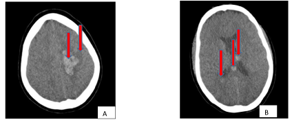
An emergency CT scan revealed a left precentral acute frontal hematoma extending to the ipsilateral caudate nucleus, with intraventricular extension in the left lateral ventricle with surrounding vasogenic edema (Figure 1).
The electrocardiogram was normal as was the chest x-ray.
The biological investigation showed:
- Hypochromic normocytic anemia at 8.8g/dl with predominantly neutrophil leukocytosis at 15300/mm3 with 13300 neutrophil/mm3, platelets at 481000/mm3.
- Hyper uremia at 2.60g/l (0.15-0.45), serum creatinemia at 205 mg/l (7-14), a blood glucose level at 0.68 g/l (0.74-1.10), SGOT = 25 IU/l (< 3), SGPT = 20 IU/l (< 32), alkaline phosphatase = 202 IU/l (98-279),Gamma GT = 123 IU/l (7-32), Na+ = 136 mmol/l (135-148), a serum potassium level 2.5 mmol/l (3.5-5.3), 99 mmol/l (99-110) chloremia, 104 mg/l (90-110) serum calcium, 30 mg/l (25-50) phosphoremia.
The abdominal ultrasound showed bilateral stage 2 kidney acute failure.
The diagnosis of left frontal hematoma by envenomation was made. The patient - received an emergency treatment with Anti-Venomous Serum (SAV), an anti-edematous (Mannitol 20 %), cerebral motor physiotherapy with speech therapy.
The kidney check-up after one week of admission, had shown a serum creatinine level of 109 mg/l compared with 205 mg/l at the admission and urea at 2.91 g/l versus 2.60 g/l at the admission. After sixteen days of hospitalization, the patient was - stable. We could not carry out the biological examinations and the cerebral scanner of control for lack of financial means. However, there has been a good clinical outcome.
Case 2
This was a 31-year-old farmer referred patient from a peripheral hospital (related to the prefecture/ bigger than peripheral) hospital located 52 km from Kara Teaching Hospital for left hemiparesis.
The patient had been bitten two weeks earlier by a viperid and taken care of at Kante district Hospital, located fifty-two kilometers from Kara Hospital. Despite the first dose of anti-venomous serum three days after the bite, the patient presented a severe anemia, which led to his transfer to Kara Teaching Hospital where he had transfusion with several additional doses of SAV. The patient’s clinical condition had been stabilized and the patient released after eight days of hospitalization B.
Two days after his return home the patient had been readmitted for sudden stroke preceded by severe headaches rated 8/10 at the EVA with nausea and vomiting. The patient was readmitted to the referred Kara CHU of the Kante Prefectural Center.
The examination on admission, three weeks after he has been bitten demonstrated
- Normal vigilance with no phasic disorder
-He had a predominantly brachio-facial left hemiparesis with segmental strength at 3/5 on the upper and 4/5 on the lower limbs, diminished osteotendinous reflexes and a Babinski sign.
- There was a left polymodal hemihypoesthesia.
- There was a minor left visuospatial hemineglect
- He had a left homonymous hemianopia
- He had a normal fundus examination
- He had no of oculomotor disorder
- The rest of the neurological examination was normal
At the cardiovascular level, the examination made it possible to note that:
- He had a blood pressure of 150/70 mmHg on the right arm and 144/71 on the left arm.
- There was a regular pulse at 88 beats per minute
-There was absence of peripheral signs of heart failure
-There were regular heart sounds at 88 beats per minute without added breath or noise.
- The examination of lungs was normal
- The rest of the physical examination.
The cerebral CT scan revealed acute right parietal cortical hematoma with obliteration of the occipital horn of ipsilateral lateral ventricle and surrounding edema (Figure 2: C and D).
Complete blood count at the admission showed an anemia with 9.8 g/dl of hemoglobin, platelets at 481000/mm3. The chest Xray, the electrocardiogram and the bottom of the eyes were normal. HIV serology was negative. The activated partial thromboplastin time was 29 seconds (25-39), the prothrombin time was 84% (70-100), the serum sodium was 135 mmol/l (135-148), the serum potassium was 3.8 mmol/l (3.5-5.3), and the plasma chloremia level was 100 mmol/l (98-110).
After ten days of hospitalization, the patient clearly recovered with a segmental force of 4 +/ 5 to the deficit members.
After ten days of hospitalization, there was a favorable outcome. We could not carry out the biological examinations and the cerebral scanner of control for lack of financial means. However, there has been a good clinical outcome.
Discussion
We report two cases of intra cerebral hematoma in two young patients with no significant past history, aged 27 and 31 years respectively. These intra cerebral hematomas complicated from a snake bite with a delay in antivenom (FAV-Afrique®) treatment. These hematomas are explained by the action of snake venom. Indeed, various disturbances of hemostasis can occur as a result of a snake bite [6]. Almost all Ophidian species responsible for serious human or fatal human envenomation are known. Venoms of these snakes are rich in proteins interfering with haemostasis, including many enzymes. These proteins can be classified in four groups according to their action (prothrombin activator, thrombin-like enzymes, factor X and factor V activators). Proteins disrupting primary haemostasis can both activate and inhibit platelets: phospholipases A2, serine proteases and metalloproteases, L-amino acido-oxidases, phosphoesterases, disintegrins, type C lectins, dendropeptins, aggregoserpentins, thrombolectins. Proteins interfering with coagulation are distinguished between procoagulant proteases and anticoagulant proteases. The venom components acting on fibrinolysis are fibrinolytic enzymes or plasminogen activators. The most common clinical consequence of these mechanisms is a local or diffuse hemorrhagic syndrome including often fatal intracranial hemorrhage [6].
However, cases of hemorrhagic stroke by envenomation are rare and reported in the literature as clinical cases [7-9]. Their interest lies in their evolution, often marked by dramatic or even fatal complications [10]. It is therefore necessary to set up a mechanism for rapid or even free management of snakebite cases in at-risk environments by the organisation of snakebite management and provision of antivenom like FAV-Afrique® which is a polyvalent snake antivenom. This antivenom is elaborated by immunisation of horses with venom from 10 different snake species among the most dangerous in Africa and belonging to Elapidae and Viperidea families. Only fragment antigen binding 2 (F(ab’)2) fragments are kept and purified. This serum is able to decrease the quantity of circulating venom and therefore its toxicity. Its use is indicated as soon as the first signs of poisoning are observed (local oedema). Twenty millimetres are administrated via intra-venous route whatever the weight of the patient. Re-administration may be performed if improvement is not sufficient. Treatment should be initiated as soon as possible but can be realized as long as the symptoms are present [11].
Conclusion
Snakebite envenomation is a neglected tropical disease that affects many millions of people worldwide, especially in rural areas. The neurovascular complications of these bites are often dramatic. Prevention by raising the awareness of target populations and better organization of our health systems will reduce the incidence and morbidity-mortality related to these envenomation by a fast and effective treatment of cases of envenomation.
- Katibi OS, Adepoju FG, Olorunsola BO, Ernest SK, Monsudi KF. Blindness and scalp haematoma in a child following a snakebite. Afri Health Sci. 2015; 15: 1041-1044. https://goo.gl/kcMNbc
- Chippaux JP. Estimate of the burden of snakebites in sub-Saharan Africa: a meta-analytic approach. Toxicon 2011; 57: 586-599. https://goo.gl/NGqQf9
- Kasturiratne A, Wickremasinghe AR, de Silva N, Gunawardena NK, Pathmeswaran A, Premaratna R, et al. The global burden of snakebite: a literature analysis and modelling based on regional estimates of envenoming and deaths. PLoS Med. 2008; 5: 218. https://goo.gl/K2JspF
- Chippaux JP. Venins de serpent et envenimations. Paris: Collection Didactiques: Edition IRD; 2002. Collection Didactiques. p.288.
- Del Brutto OH, Del Brutto VJ. Neurological complications of venomous snake bites: a review. Acta Neurol Scand: 2012; 125: 363-372. https://goo.gl/8Mx5sY
- Larreche S, Mion G, Goyffon M. Haemostasis disorders caused by snake venoms. Ann Fr Anesth Reanim. 2008; 27: 302-309. https://goo.gl/Bd4LBK
- Pinho FM, Burdmann EA. Fatal cerebral hemorrhage and acute renal failure after young Bothrops jararacussu snake bite. Ren Fail. 2001; 23: 269-277. https://goo.gl/AnuDfm
- Berling I, Isbister GK. Hematologic effects and complications of snake envenoming. Transfus Med Rev. 2015; 29: 82-89. https://goo.gl/Pa5yPW
- Mosquera A, Drover LA, Tafur A, Del Brutto OH. Stroke following Bothrops spp. snakebite. Neurology. 2003; 60: 1577-1580. https://goo.gl/ssqdgZ
- Ghezala HB, Snouda S. Hemorrhagic stroke following a fatal envenomation by a horned viper in Tunisia. Pan Afr Med J. 2015; 21: 156. https://goo.gl/6vQjsP
- Wolf A, Mazenot C, Spadoni S, Calvet F, Demoncheaux JP. FAV-Africa®: a polyvalent antivenom serum used in Africa and Europe. Med Trop (Mars). 2011; 71: 537-540. https://goo.gl/VpR3MP


Sign up for Article Alerts