Malignant Peripheral Nerve Sheath Tumor Rare Neurological Isolated Neoplasm?
Idrissi Serhrouchni K1*, Hammas N2, El Fatemi H2, Chbani L2, Harmouch T2 and Amarti A2
1Department of Pathology, Oncology Center, Tangier, 90000, Morocco
2Department of Pathology, Hassan II University Hospital, Fez, 30000, Morocco
*Address for Correspondence: Idrissi Serhrouchni Karima, Department of Pathology, Regional Center of Oncology, Tangier, 90000, Morocco, Tel: +212-661-444-751; E-mail: [email protected]
Submitted: 25 October 2018; Approved: 05 January 2019; Published: 08 January 2019
Citation this article: Idrissi Serhrouchni K, Hammas N, El Fatemi H, Chbani L, Harmouch T, et al. Malignant Peripheral Nerve Sheath Tumor Rare Neurological Isolated Neoplasm. Int J Neurol Dis. 2019;3(1): 001-003.
Copyright: © 2019 Idrissi Serhrouchni K, et al. This is an open access article distributed under the Creative Commons Attribution License, which permits unrestricted use, distribution, and reproduction in any medium, provided the original work is properly cited
Keywords: Malignant Peripheral Nerve Sheath Tumor (MPNST); Neurofibromatosis; Protein S100
Download Fulltext PDF
Primary malignant schwannoma is a rare neoplasm of nerve sheath origin. It is a cancer of the connective tissue surrounding nerves. Given its origin and behavior it is classified as sarcoma. The estimated incidence of Malignant Peripheral Nerve Sheath Tumor (MPNST) in general population is 0.001% and in patients with Neurofibromatosis 1 (NF-1) is 2-5%. It is an uncommon spindle cell sarcoma accounting for approximately 5% of all soft-tissue sarcoma. They are highly aggressive and occur in the second or third decade. This neoplasm usually affects the extremities. There is strong association between MPNSTs and neurofibromatosis (NF-1) and previous irradiation. We present the case of a 61-year-old woman manifesting with recurrent sciatalgy near for the fourth and fifth lumbar vertebral bodies. She underwent resection of a mass at the L4-5 level that was subsequently recognized as a malignant peripheral nerve sheath tumor.
Introduction
Malignant Peripheral Nerve Sheath Tumor (MPNST) is extremely rare entity. However, it is of diagnostic importance because it is one of the most aggressive soft-tissue sarcomas. It is also called as neurofibrosarcoma as it arises from the neuroectodermal lining of the peripheral nerves. MPNSTs are often associated with NF1 in 30-50% and with previous irradiation in 4-11% while 0.001% arise de novo in the general population [1,2]. They often affect the head, trunk, and extremities; they lead to poor overall survival [3].
Case Report
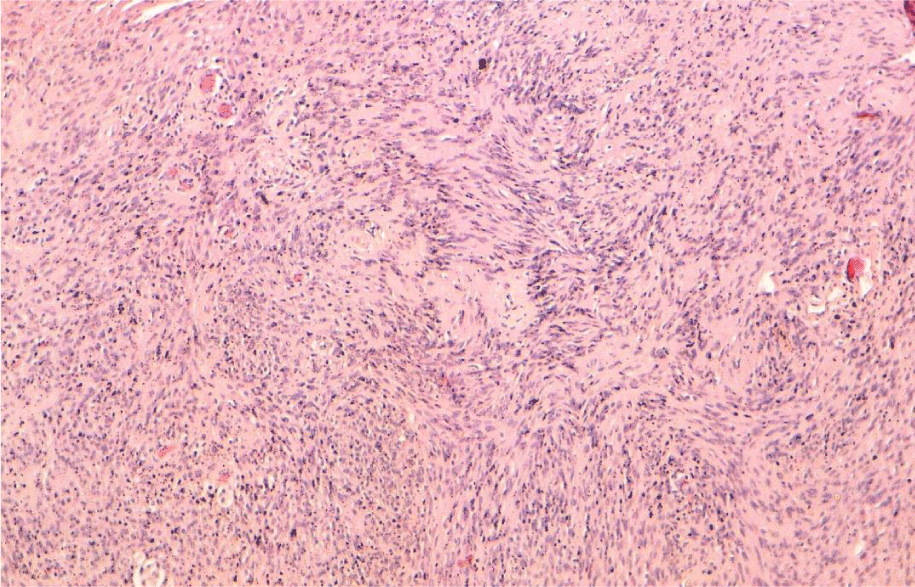
A 61- year-old female patient presented with history of persistent pain in the lowest segment of the lumbar region and sciatalgy since two years, without favourable evolution using medical treatment. Physical examination revealed a sensibility on her lumbar vertebrae near for the L4-L5. Nerve function and the remainder of general examination were normal without skin, skeletal or ocular anomalies. Computed Tomography (CT) scan showed a medullar necrotic rounded mass lesion at the L4-L5 level. Histopathological examination of the specimen revealed a tumor composed of spindle shaped cells with elongated hyperchromatic nuclei, arranged in long and short fascicles. Cells were arranged around hyaline bands in the form of cords and at places they were arranged in whorls (Figure 1). Some of the tumor cells had marked nuclear pleomorphism. The stroma showed extensive areas of necrosis and hemorrhage. The blood vessels showed hyalinized walls. Mitotic figures were more than five per high-power field (Figure 2). Immunohistochemically, the tumor cells showed diffuse and strong positivity for S-100 (Figure 3). However, they were negative for Pan-cytokeratin (AE1/ AE3), Desmin, and Human Melanoma Black (HMB) 45. Based on this data, a diagnosis of MPNST was made.
Discussion
MPNSTs are malignant tumors developing from cells of peripheral nerve tissue [2]. They are also called neurofibrosarcomas, malignant schwannoma or neurogenic sarcoma and account for approximately 5-10% of all soft-tissue sarcomas and about one-fourth to one-half occur in the setting of NF-1 [4]. The MPNST is typically a disease of adult life, as most tumors occur in patients of 20-50 years of age [5]. It does not exhibit any sex differences [4]. The age of onset tends to be lower in cases associated with NF-1 [6].
More than 50% patients with MPNSTs suffer from NF-1, our case is not integrated in the syndrome of NF-1 it presents a form of an isolated neoplasm.
The most common anatomical sites for MPNST include extremities, head and neck region and trunk. Evans et al showed in their study about 32 cases of MPNST with Twelve (38%) occurred in deep locations with no superficial signs of a plexiform tumor. Many others occurred on major nerve routes such as the brachial plexus or sciatic nerve. Few tumors occurred at extremities [7].
CT findings of MPNSTs usually include a large heterogeneous mass with occasional bony destruction. At MRI, The signal intensity of the tumors on T1WI is equal to or slightly greater than that of muscles. T2WI reveals high signal intensity and heterogeneous enhancement with partial central necrosis after contrast enhancement [8]. The most accurate radiographic evaluation of MPNST uses a combination of PET along with CT or MRI [9]. However diagnosis could not be made only on the basis of imaging studies, but it needs histological confirmation.
Histopathologically, MPNSTs are characterized by atypical spindle-shaped cells with nuclear pleomorphism, frequent mitosis, and a high proliferative index. Tumor cells show S-100 immunopositivity, implying nervous origin, and vimentin immunopositivity, implying a mesenchymal origin-sarcoma [10]. The pathologic diagnoses of peripheral nerve sheath tumors with atypia represent a histologic continuum, and include neurofibroma with atypical features, low-grade MPNST, and high-grade MPNST [9].
MPNSTs have to be differentiated on the one hand from neurofibromas sand schwannomas and on the other form the others spindle cell sarcomas. Schwannomas are encapsulated tumors of nerve sheath with palissading form of nuclei, without mitotic activity or nuclear atypia. MPNSTs are hyper-cellular and show pleomorphism and mtotic figures. Low grade MPNSTs arising from neurofibroma is diagnosed when there is generalized nuclear atypia, increased cellularity and unusually low-levels of mitotic activity [2].
Malignant transformation is extremely rare in schwannomas, with fewer than 20 reported cases. Unlike neurofibromas, which most often give rise to conventional spindle cell malignant peripheral nerve sheath tumors. Sarcomas arising in schwannomas frequently show epithelioid morphology, including epithelioid malignant peripheral nerve sheath tumor and epithelioid angiosarcoma [11].
The development of neurofibromas and their subsequent progression to become MPNSTs involves a sequential series of tumor suppressor mutations. Deletions and other mutations that result in loss of function of the TP53 tumor suppressor gene are some of the more common abnormalities found in MPNSTs. Wide surgical excision of tumor and radical dissection is the treatment of choice for MPNSTs arising in trunk. The role of chemotherapy in MPNST treatment remains controversial.
The most common metastatic site of MPNST is the lung. According to Wise et al. [12] the prognosis of MPNSTs is poor and have 5 year survival rate of 20-50%. Large tumor size ( > 5 cm), presence of neurofibromatosis, and total resection are most important prognostic indicators [13]. Histologic findings such as cellularity, pleomorphism, mitotic activity, and size also contribute in assessing the prognosis [14]. Wong et al. [14,15] reported that adjuvant irradiation ( > 60 Gy) and inclusion of intraoperative electron irradiation were associated with better local disease control. Our patient received routine adjuvant chemotherapy after complete resection without complications or recurrence.
Conclusion
Our case point out that in case of persistent pain and resistant to medical therapy, associated with alterations of sensitivity, neoplastic causes including the tumors of the nervous system should be considered. Although malignant medullary tumors are rare, MPNSTs should be included in the differential diagnosis of the nervous system tumors. Complete resection and administration of routine adjuvant chemotherapy are the treatment of choice.
- Gogate BP, Anand M, Deshmukh SD, Purandare SN. Malignant peripheral nerve sheath tumor of facial nerve: Presenting as parotid mass. J Oral Maxillofac Pathol. 2013; 17: 129-131. https://goo.gl/jRfA5H
- Neville H, Corpron C, Blakely ML, Andrassy R. Pediatric neurofibrosarcoma. J Pediatr Surg. 2003; 38: 343-346. https://goo.gl/QCp8Ge
- Tucker T, Wolkenstein P, Revuz J, Zeller J, Friedman JM. Association between benign and malignant peripheral nerve sheath tumors in NF1. Neurology. 2005; 65: 205-211. https://goo.gl/zhMbq2
- Richards JB, Marotti J, DeCamp MM, Roberts DH. Thoracic malignant peripheral nerve sheath tumor: a case report and literature review. Clin Pulm Med. 2012; 19: 298-301. https://goo.gl/oZRh9u
- Weiss SW, Goldblum JR. Malignant tumors of the peripheral nerves. In: Weiss SW, Goldblum JR, editors. Enzinger and Weiss's Soft Tissue Tumors. 2nd ed. Philadelphia: Mosby. 1998; 903-944.
- Hruban RH, Shiu MH, Senie RT, Woodruff JM. Malignant peripheral nerve sheath tumors of the buttock and lower extremity. A study of 43 cases. Cancer. 1990; 66: 1253-1265. https://goo.gl/FpPS7z
- Evans DG, Baser ME, McGaughran J, Sharif S, Howard E, Moran A. Malignant peripheral nerve sheath tumours in neurofibromatosis 1. J Med Genet. 2002; 39: 311-314. https://goo.gl/iH8F36
- Tateishi U, Gladish GW, Kusumoto M, Hasegawa T, Yokoyama R, Tsuchiya R, et al. Chest wall tumors: radiologic findings and pathologic correlation: part 2. Malignant tumors. Radiographics. 2003; 23: 1491-508. https://goo.gl/hyMdYf
- Aaron WJ, Elizabeth S, Arun S, Sarah MD, Fritz CE. Malignant peripheral nerve sheath tumor. Surg Oncol Clin N Am. 2016; 25: 789-802.
- Lee CF, Luo JW, Liu CJ. Undifferentiated sarcoma of the mandible: a case report. Taiwan J Oral Surg 2009; 20: 263-271.
- Carter JM, O'Hara C, Dundas G, Gilchrist D, Collins MS, Eaton K, et al. Epithelioid malignant peripheral nerve sheath tumor arising in a schwannoma, in a patient with “neuroblastoma-like” schwannomatosis and a novel germline smarcb1 mutation. Am J Surg Pathol. 2012; 36: 154-160. https://goo.gl/Krg5Lq
- Wise JB, Patel SG, Shah JP. Management issues in massive pediatric facial plexiform neurofibroma with neurofibromatosis type 1. Head Neck. 2002; 24: 207-211. https://goo.gl/C8h175
- Ducatman BS, Scheithauer BW, Piepgras DG, Reiman HM, Ilstrup DM. Malignant peripheral nerve sheath tumors. A clinicopathologic study of 120 cases. Cancer. 1986; 57: 2006-2021. https://goo.gl/Gj5AGj
- Colreavy MP, Lacy PD, Hughes J, Bouchier-Hayes D, Brennan P, O’Dwyer AJ, et al. Head and neck schwannomas: A 10 year review. J Laryngol Otol. 2000; 114: 119-124. https://goo.gl/xcKYQ6
- Wong WW, Hirose T, Scheithauer BW, Schild SE, Gunderson LL. Malignant peripheral nerve sheath tumor: analysis of treatment outcome. Int J Radiat Oncol Biol Phys. 1998; 42: 351-360. https://goo.gl/SyTm8u
- Hsu CC, Huang TW, Hsu JY, Shin N, Chang H. Malignant peripheral nerve sheath tumor of the chest wall associated with neurofibromatosis: a case report. J Thorac Dis. 2013; 5: 78-82. https://goo.gl/hKpKZt


Sign up for Article Alerts