Petersen’s Hernia after Laparoscopic Roux-En-Y Gastric Bypass Presenting in Second Trimester Pregnancy?
Michael B. Goldberg*, Ali Tavakkoli, Malcolm K. Robinson
Brigham and Women’s Hospital, Boston, MA
*Address for Correspondence: Michael B. Goldberg, Brigham and Women’s Hospital, Boston; E-mail: [email protected]
Submitted: 01 May 2017; Approved: 17 May 2017; Published: 22 May 2017
Citation this article: Goldberg MB, Tavakkoli A, Robinson MK. Petersen’s Hernia after Laparoscopic Roux-En-Y Gastric Bypass Presenting in Second Trimester Pregnancy. SRL Reprod Med Gynecol. 2017;3(1): 020-023.
Copyright: © 2017 Goldberg MB, et al. This is an open access article distributed under the Creative Commons Attribution License, which permits unrestricted use, distribution, and reproduction in any medium, provided the original work is properly cited
Download Fulltext PDF
Introduction
One third of all women in the United States are obese [1], and approximately one-fifth of the population is obese in pregnancy [2]. Obesity during pregnancy caries multiple risks to the fetus and the mother, including fetal macrosomia, prematurity, miscarriage, maternal hypertension, and gestational diabetes [1,3].
Bariatric surgery is gaining popularity to treat obesity and its related comorbidities, especially among women of childbearing age. While bariatric surgery is not a treatment for infertility, women may experience an increase in fertility postoperatively as conditions such as Polycystic Ovarian Syndrome (PCOS) resolve with weight loss [4]. Furthermore, bariatric surgery may be an attractive option to women of childbearing age to get to a healthy weight before planning to conceive in order to mitigate obesity-related risks to the fetus. Controversy exists regarding the time interval that patients must wait after bariatric surgery to safely conceive. Generally, women are advised to wait 12-24 months after surgery until weight and nutritional status stabilize [1,4].
Internal hernia after gastric bypass is a serious complication which requires prompt surgical therapy to prevent bowel necrosis. The incidence of internal hernia after Roux-en-Y Gastric Bypass (RYGB) ranges from 0.2 to 11% [5], and is associated with a mortality rate of 1.6% [6]. The evaluation of pregnant patients with a history of bariatric surgery requires special consideration — abdominal pain, nausea, and vomiting can signify a potential surgical emergency due to the gastric bypass or be related to the pregnancy. We report two cases of internal hernias through Petersen’s defect presenting in the second trimester of pregnancy. Both patients underwent operative repair without complications.
Case 1
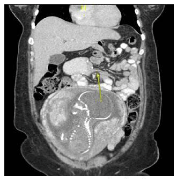
A 27-year-old female in her 26th week of pregnancy presented to the emergency department with one day of colicky epigastric pain, nausea, and vomiting without changes in bowel habits. She had undergone laparoscopic RYGB (antecolic, antegastric) 18 months previously with a weight loss of 123 pounds (preoperative BMI 49 decreased to 28). On presentation, the patient appeared uncomfortable in bed. Her abdominal exam was gravid with mild epigastric tenderness and no peritoneal signs. The patient was initially evaluated by the obstetric team, where a bedside fetal ultrasound was performed and did not reveal any cause of her pain. The patient’s vital signs and laboratory studies were normal, including a White Blood Cell count (WBC) of 7.4 K / ul. After discussing the risks and benefits of abdominal imaging, we proceeded with a Computed Tomographic (CT) scan of the abdomen and pelvis with oral and intravenous contrast. Imaging revealed a prominent small bowel loop with swirling of mesenteric fat and vessels in the left upper quadrant of the abdomen behind the Roux limb concerning for an internal hernia (Figure 1). There was no complete bowel obstruction as oral contrast passed to distal small bowel loops. An exploratory laparoscopy was recommended and the patient was brought to the operating room emergently. Preoperatively, antibiotics and chemical thromboprophylaxis were administered. The patient was positioned with a roll under her right side to prevent compression of the Inferior Vena Cava (IVC) during the surgery. The fetus was monitored throughout the procedure via ultrasonic heart rate monitoring with a probe placed in the lower aspect of the abdomen underneath the surgical drapes. The abdomen was entered using an open hasson technique above the umbilicus and insufflated with carbon dioxide to a pressure of 12 mmhg; this is a lower pressure than normally used to help maintain adequate venous return. A total of four ports were used. The roux limb was examined and run distally which revealed internal herniation of the distal roux limb, jejunojejunostomy, and proximal portion of the common channel through Petersen’s defect. Once the bowel was reduced from this hernia, the RYGB anatomy was restored and we were able to examine the bowel from the ligament of Treitz distally to the terminal ileum. The bowel was proximally dilated and distally decompressed, and it was all viable. The Petersen’s defect was closed using running silk suture. Throughout our laparoscopic procedure, the fetal heart rate was normal. The patient tolerated the procedure and was extubated and transferred to the post-anaesthesia care unit in stable condition. A clear liquid diet was started on postoperative day one and the patient was discharged home on postoperative day four. There were no fetal or maternal complications. The patient underwent planned cesarean section at 39 weeks with delivery of a healthy baby boy.
Case 2
A 35-year-old female in her 25th week of pregnancy presented to the emergency department with abdominal pain, nausea, and vomiting for three days. She described her pain as left-sided and crampy. Although the patient experienced emesis throughout her pregnancy, it was more frequent during this episode. She had undergone laparoscopic RYGB (antecolic, antegastric) 13 months prior to presentation at an outside hospital, and she reported weight loss of over 80 pounds. The patient presented with a BMI of 22 from a preoperative BMI of 33.5. Vital signs were normal on presentation and laboratory studies revealed a lactic acid level of 3.6 mmol / L and a WBC of 21.6 K/ul. Electrolytes and hepatic function tests were normal. Abdominal exam was benign without any tenderness or peritoneal signs. A CT scan of the abdomen and pelvis with oral and intravenous contrast revealed passage of oral contrast into distal small bowel loops, however there was swirling of the mesenteric vasculature and mesenteric congestion concerning for an internal hernia (Figure 2). Given the patients concerning laboratory values and imaging, exploratory laparotomy was recommended and the patient was brought to the operating room emergently. Preoperatively, antibiotics and chemical thromboprophylaxis were administered. The patient was positioned with a roll under her right side and the fetus was monitored throughout the procedure. The patient’s abdomen was entered via upper midline incision and the bowel was inspected, beginning at the terminal ileum and exploring in a retrograde fashion back to the jejunojejunostomy. An internal hernia in the Petersen’s space was encountered with cyanotic bowel in the common limb. Once the bowel was reduced it appeared viable after several minutes. The Petersen’s defect was closed using three interrupted silk sutures. After a final exploration, the abdomen was closed. Throughout our exploration, the fetal heart rate was normal. Postoperatively, the patient’s lactate and WBC decreased to normal values. She tolerated a clear liquid diet on postoperative day one and was discharged home on postoperative day three. There were no fetal or maternal complications. The patient underwent spontaneous vaginal delivery at 40 weeks with delivery of a healthy baby girl.
Discussion
Bariatric surgery is gaining favour to treat obesity and its related conditions, with the majority of patients being women of childbearing age. Even in women with regularly occurring menstrual cycles, obesity is associated with decreased fertility due to oligo- and an ovulation and PCOS, which may not cause any change in the menstrual cycle. Obesity during pregnancy is associated with increased risk to the fetus (prematurity, macrosomia, dystocia, neonatal death), to the mother (hypertension, gestational diabetes, thrombosis), and during delivery (difficult delivery, birth injury, anaesthetic complications, infection, postpartum haemorrhage, and maternal mortality) [1]. Fertility and fetal complications improve after weight loss from bariatric surgery. Some studies suggest bariatric surgery before pregnancy is associated with reduced prevalence of fetal anomalies, maternal diabetes and hypertension [3]. However, data on fetal outcomes after bariatric surgery is mixed, as some studies have found increased risk of Small-for-Gestational-Age (SGA) infants and shorter gestation [7]. The time to wait after surgery to safely conceive is debated and largely based on expert opinion. While most surgeons advise patients to avoid pregnancy until weight and nutritional status are sable (usually after 12-24 months), the American College of Gynecologists recommends avoiding pregnancy for a minimum of two years after a bariatric operation [4]. Fetal outcomes in mothers who conceive less than two years after bariatric surgery may be associated with higher risks of prematurity, NICU admission, and SGA status compared to mothers who wait longer [3].
Increasingly, surgeons are faced with the difficult clinical dilemma of a pregnant post-bariatric patient with abdominal complaints. While abdominal pain during pregnancy can be difficult to work up, prompt diagnosis is necessary to prevent adverse maternal and fetal outcomes [8]. Even in pregnant patients without a history of RYGB, it is important to consider adhesive small bowel obstruction if the patient has a history of previous abdominal operations. In patients without peritonitis or hemodynamic instability in whom diagnosis may be unclear, we recommend abdominal imaging with a CT scan with oral and intravenous contrast when feasible. While ultrasound is safe and commonly performed in pregnant patients, this imaging modality is typically not useful to diagnose an internal hernia. Radiation may cause teratogenesis of the nervous system between 10 and 17 weeks gestation and increase risk of developing childhood hematologic malignancy by 0.06 % per 1 rad delivered to the fetus. Depending on institutional protocols an abdominopelvic CT scan can reach 5 rads, which is considered safe. It is recommended that the total cumulative dose of radiation to a fetus during pregnancy be less than 5 - 10 rads [9]. An alternative to CT scanning is Magnetic Resonance (MR) imaging. This modality can be performed at any stage in pregnancy without risks of ionizing radiation and must be done without IV gadolinium contrast. While risks to the fetus are reduced with MR, the ease of obtaining and interpreting these images is limited by institutional availability and may lead to a delay in treatment.
Once internal hernia is suspected, operative intervention should not be delayed as bowel resection is associated with adverse maternal and fetal outcomes10. It has been suggested that the risk of internal hernia increases as pregnancy progresses because the abdominal viscera is displaced by the gravid uterus [11]. While we favour cross sectional imaging in the work up of the pregnant bariatric patient with abdominal pain, high clinical concern mandates operative exploration even without imaging. Similarly, in the pregnant non-bariatric patient with bowel obstruction and concern for bowel compromise, we recommend maintaining a low threshold for operative intervention.
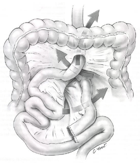
Small bowel obstruction occurs from internal hernia in 0.2 – 11 % of all patients [5]. During antecolic gastric bypass, internal hernias can occur at the mesenteric defect at the jejunojejunostomy and at Petersen’s space—the open space between the mesentery of the alimentary (roux) limb and the mesentery of the transverse colon. When RYGB is done in a retrocolic configuration, the space created in the transverse colon mesentery adds another potential site for internal herniation (Figure 3). It is believed that internal hernias are more common after laparoscopic gastric bypass compared with open surgery due to decreased adhesion formation and lack of bowel fixation. Most recent data supports the need for closure of all mesenteric defects to reduce the risk of internal hernias [5,12,13]. While this risk is inherent to RYGB, closing the defects during the index operation is currently the only way to reduce future risk. The authors of this case report favour closure of mesenteric defects with a non absorbable braided suture during gastric bypass.
Based on surgeon comfort and preference, exploration can be done laparoscopically or via midline laparotomy. Laparoscopic exploration can be safely performed during any trimester of pregnancy without increased risk to the mother or fetus [9]. In our second case, an open approach was chosen given the relatively low BMI of 22 and gravid uterus raising concern for adequate domain within which to operate with a reduced working space. While it would have been reasonable to approach this case laparoscopically at first, it is important to underscore the need for prompt intervention based on surgeon comfort. It is imperative to involve the obstetric team in the care of these complex patients and at a minimum, fetal heart monitoring should occur pre- and postoperatively. To limit compression on the IVC, patients should be positioned with the right side up. During laparoscopic exploration, we favour entering the abdomen via open hasson technique to avoid damage to the gravid uterus or any displaced abdominal viscera. Operative exploration mandates inspection of the entirety of the small bowel and all mesenteric defects.
We present two successful outcomes after laparoscopic and open operative exploration, reduction and repair of Petersen’s hernias in pregnant patients. It is imperative to maintain a high clinical suspicion for this potentially lethal surgical emergency.
- Narayanan RP, Syed AA. Pregnancy following bariatric surgery-medical complications and management. Obes Surg. 2016; 26: 2523-2529. https://goo.gl/XjTb4z
- Willis K, Lieberman N, Sheiner E. Pregnancy and neonatal outcome after bariatric surgery. Best Pract Res Clin Obstet Gynaecol. 2015; 29: 133-144. https://goo.gl/MC41rF
- Parent B, Martopullo I, Weiss NS, Khandelwal S, Fay EE, Rowhani-Rahbar A. Bariatric surgery in women of childbearing age, timing between an operation and birth, and associated perinatal complications. JAMA Surg. 2017; 152: 1-8. https://goo.gl/O23kOz
- American College of Obstetricians and Gynecologists. ACOG practice bulletin No. 105: bariatric surgery and pregnancy. Obstet Gynecol. 2009; 113: 1405-1413. https://goo.gl/3i8P3x
- Koppman JS, Li C, Gandsas A. Small bowel obstruction after laparoscopic Roux-en-Y gastric bypass: a review of 9,527 patients. J Am Coll Surg. 2008; 206: 571-584. https://goo.gl/BA98md
- Fabozzi M, Brachet Contul R, Millo P, Allieta R. Intestinal infarction by internal hernia in Petersen's space after laparoscopic gastric bypass. World J Gastroenterol. 2014; 20: 16349-16354. https://goo.gl/abESfw
- Johansson K, Cnattingius S, Näslund I, Roos N, Trolle Lagerros Y, Granath F, et al. Outcomes of pregnancy after bariatric surgery. N Engl J Med. 2015; 372: 814-824. https://goo.gl/ovtdt3
- Moore KA, Ouyang DW, Whang EE. Maternal and fetal deaths after gastric bypass surgery for morbid obesity. N Engl J Med. 2004; 351: 721-722. https://goo.gl/PFydK0
- Pearl J, Price R, Richardson W, Fanelli R, Society of American Gastrointestinal Endoscopic Surgeons. Guidelines for diagnosis, treatment, and use of laparoscopy for surgical problems during pregnancy. Surg Endosc. 2011; 25: 3479-3492. https://goo.gl/CFEOQK
- Efthimiou E, Stein L, Court O, Christou N. Internal hernia after gastric bypass surgery during middle trimester pregnancy resulting in fetal loss: risk of internal hernia never ends. Surg Obes Relat Dis. 2009; 5: 378-380. https://goo.gl/r3VD7z
- Gudbrand C, Andreasen LA, Boilesen AE. Internal hernia in pregnant women after gastric bypass: a retrospective Register-based cohort study. Obes Surg. 2015; 25: 2257-2262. https://goo.gl/YP9VZ4
- Stenberg E, Szabo E, Ottosson J, Näslund I. Outcomes of laparoscopic gastric bypass in a randomized clinical trial compared with a concurrent national database. Br J Surg. 2017; 104: 562-569. https://goo.gl/dNEBSV
- Aghajani E, Nergaard BJ, Leifson BG, Hedenbro J, Gislason H. The mesenteric defects in laparoscopic Roux-en-Y gastric bypass: 5 years follow-up of non-closure versus closure using the stapler technique. Surg Endosc. 2017. https://goo.gl/bGb2WE
- Gagne DJ, DeVoogd K, Rutkoski JD, Papasavas PK, Urbandt JE. Laparoscopic repair of internal hernia during pregnancy after Roux-en-Y gastric bypass. Surg Obes Relat Dis. 2010; 6: 88-92. https://goo.gl/t04bYb
- Gruetter F, Kraljevic M, Nebiker CA, Delko T. Internal hernia in late pregnancy after laparoscopic Roux-en-Y gastric bypass. BMJ Case Rep. 2014. https://goo.gl/R8H1hM


Sign up for Article Alerts