A Case Report of a Rare Finding of Fetal Anterior Neck Mass
Haile G Mekuria1*, Helina A Abebe2 and Hilkiah K Suga2
1Department of Obstetrics and Gynecology, Myungsung Christian Medical Center, Addis Ababa, Ethiopia
2Senior Medical Student, Myungsung Medical College, Addis Ababa, Ethiopia
*Address for Correspondence: Haile G. Mekuria, Department of Obstetrics and Gynecology, Myungsung Christian Medical Center, Addis Ababa, Ethiopia, E-mail: [email protected]
Submitted: 20 April 2019; Approved: 14 May 2019; Published: 16 May 2019
Citation this article: Mekuria HG, Abebe HA, Suga HK. A Case Report of a Rare Finding of Fetal Anterior Neck Mass. Int J Reprod Med Gynecol. 2019;5(1): 016-018.
Copyright: © 2019 Mekuria HG, et al. This is an open access article distributed under the Creative Commons Attribution License, which permits unrestricted use, distribution, and reproduction in any medium, provided the original work is properly cited
Keywords: Fetal anterior neck teratoma; Cystic mass; Polyhydramnios
Download Fulltext PDF
Introduction
Fetal anterior neck teratomas are tumors which arise from the three blastomeric layers - ectoderm, endoderm and mesoderm. It occurs when the totipotent germ cells are out of control of primary organizers [1,2]. The histologic features may include cystic and solid areas with organoid patterns and it may include mature or immature cells [1]. Even though the most common area of occurrence is at sacrococcgeal area it can also occur in other body parts [1,3]. In this case report we presented one of the rare place of teratoma - anterior fetal neck teratoma.
Case Report
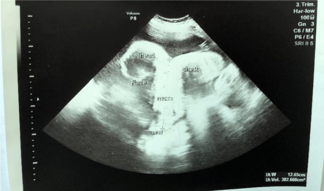
A 25-years-old PIAI patient had a smooth course of pregnancy until 21 weeks of gestation when an anterior neck mass with cystic solid complex found on the fetus during routine ultrasound screening. The mass measured 7x4 cm. Amniocentesis was done and the result was normal. Karyotype was 46 XY and trisomy 18, 21 and 13 were negative. At 27th weeks gestational age, the patient presented with preterm labor. The ultrasound showed polyhydramnios. The size of the mass reached 10x5 cm and the EFW was 1.0 kg (figure 1). The patient has no family history of similar condition. She was put on tocolytics but the labor couldn’t stop. Therefore, the family was counselled and they opt for vaginal delivery and refused cesarean delivery.
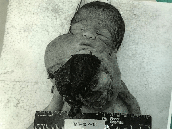
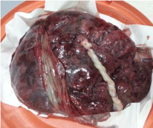
The outcome was 1.3 kg female neonate still birth with breech delivery. The placenta weighs 800gm. Fetus had anterior neck mass measuring 15x8x9 cm and extending to the right side (figure 2). The mass is partially cystic and partially friable solid pinkish white tumor with focal hemorrhage and anterior neck ulceration 9x9 cm. The APGAR score was 0.
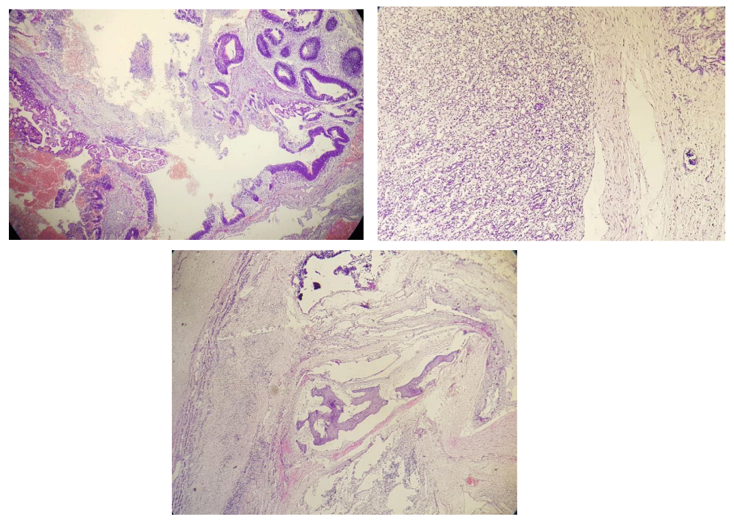
The pathology result showed section mixture of immature primitive neuroepithelial elements, choroids plexus, cartilage, bone, respiratory epithelium, skin, appendages, fibrous connective and endodermal mucinous elements (figure 3). The conclusion of the pathology report was immature teratoma partially cystic, probably thyroid origin compressing airway with ulceration.
Discussion
Fetal neck mass is a rare finding, contributes to 3-5% of all teratomas in children. There is no known factor that affects its prevalence and no association was found between fetal neck mass and Sex, race and maternal age [3]. In the majority of the cases, the mass is partly solid and partly cystic [5]. Our case also has similar finding.
Fetal Neck teratoma is formed at early gestational age and increase in size with the corresponding fetal growth. The growth of the mass causes compression of the esophagus and results decreased swallowing. This leads to polyhydramnios [3]. In addition, associated compression of the trachea also leads to tracheomalacia that may increase the post-operative morbidity [3]. Similarly, the size of the tumor in our case has grown in 6 weeks - from 7x4 at 21 weeks to 10x5 at 27 weeks. There was an associated polyhydraminos at 27th week GA which possibly led her to develop preterm labor.
Fetal anterior neck teratomas are usually associated with congenital malformations such as imperforate anus, chondrodystrophia fetalis, hypoplastic left ventricle with pulmonary hypoplasia, cystic fibrosis, absence of corpus callosum, and rarely arachanoid cyst [3]. Unlike many reported Fetal Anterior Neck Mass (FANM) cases, no malformation was found on our case.
Prenatal diagnosis of the fetal neck teratoma can be made by ultrasound examination [4]. MRI is also helpful to get a more comprehensive field of view [2]. Early diagnosis of FANM is very important because it helps to consult pediatric surgeons earlier and plan the place of delivery, the resuscitative measures that will be taken after delivery [4]. Radiological differential diagnoses of teratoma are lymphangioma, venous malformations with phleboliths, dermoid, neurenteric cysts, thornwalds cyst, and basal meningocele. [1]
The treatment of choice is surgical resection of the mass [4,5]. Ex-Utero Intrapartum Therapy (EXIT) procedure is used to secure the fetal airway before the complete delivery of the fetus [2]. In this procedure only the head and neck of the fetus will be delivered through C-section without halting the uteroplacental circulation, giving time for intubation or performing tracheostomy. After securing the airway either by intubation or tracheostomy, the fetus will be delivered completely and will be ready for resection of the mass [2].
Successful EXIT procedure needs meticulous planning of each and every steps which involves multiple disciplines such as medical ethicists, radiologists, obstetric anesthesiologists and obstetricians, pediatric surgeons and neonatologists [2]. But not all anterior neck teratoma cases don’t need EXIT procedure. Using perinatal fetoscopy helps us to identify accessible airways as a result minimize the number of cases that need EXIT procedure [6].
The prognosis of this condition depends on the time taken to stabilize and resect the tumor, degree of maturity of tissues and completeness of resection [3,4]. The long term survival and prognosis can be influenced by malignant transformation and metastasis of the tumor.
Conclusion and Recommendations
Fetal anterior neck teratoma is a rare congenital malformation which can be prenatally diagnosed by ultrasound or MRI. The tumor can obstruct the airway leading to breathing impairment after delivery. It is also commonly associated with polyhydramnios. The management is resection of the tumor after securing the airway by performing EXIT procedure. Because there are only very few studies available, the authors recommend more studies to be done on the importance of perinatal fetoscopy in determining the necessity of EXIT procedure in fetal neck teratoma cases.
Acknowledgment
The authors would like to thank our hospital, Myungsung Christian Medical Center and Dr. Selam Tadesse for their support.
- Chakravarti A, Shashidhar TB, Naglot S, Sahni JK. Head and Neck Teratomas in Children: A Case Series. Indian J Otolaryngol Head Neck Surg. 2011; 63: 193-197. https://bit.ly/2E4W9g3
- Manjiri K, Suzanne E, Theodore J, Jonathan P, Edith C. EXIT Procedure: Technique and Indications with Prenatal Imaging Parameters for Assessment of Airway Patency. RadioGraphics. 2011; 31: 511-526. https://bit.ly/2VE2dXM
- Manoj K, Pradeep S, Ashwin A, Rikki S, Samita G, Kohli G, et al. A Huge Immature Cervical Teratoma; Antenatal Diagnosis, and its Management - An Unusual Entity. J Clin Neonatol. 2013; 2: 42-45. https://bit.ly/2LGJKFe
- Shetty KJ, Kishan Prasad HL, Rai S, Kumar YS, Bhat S, Sajjan N, et al. Unusual presentation of immature teratoma of the neck: A rare case report. J Can Res Ther. 2015; 11: 647. https://bit.ly/2HjsxNT
- Jamal AA, Mansoor I, Altaf FJ, Rayes O. Clinicopathological Features of Immature Neck Teratoma. Ann Saudi Med. 2001; 21: 77-79. https://bit.ly/2Hpzyf4 Karen B, Julie F, Jan D, Pierre R, Greet H, Luc De Catte, et al. Long term outcome of pre- and perinatal management of congenital head and neck tumors and malformations. Int J Pediatr Otorhinolaryngol. 2019; 121: 164-172. https://bit.ly/2HphGB1


Sign up for Article Alerts