Endometrial Estrogen and Progesterone Receptor Expression in Women with Abnormal Uterine Bleeding in the Reproductive Age
Ahmed M. Mostafa1*, Nashwa Elsaid2, Ragaa A. Fawzy3 and Alaa Elfeky2
1Dar Elshefa Hospital
2Gynecology and Obstetrics department, Ain Shams University, Egypt
3Pathology department, Ain Shams University, Egypt
*Address for Correspondence: Ahmed M. Mostafa, Gynecology and Obstetrics department, Ain Shams University, Abbassia, Cairo, Egypt, Tel: +010-922-227-72; E-mail: [email protected]
Submitted: 10 July 2018; Approved: 30 July 2018; Published: 02 August 2018
Citation this article: Mostafa AM, Elsaid N, Fawzy RA, Elfeky A. Endometrial Estrogen and Progesterone Receptor Expression in Women with Abnormal Uterine Bleeding in the Reproductive Age. Int J Reprod Med Gynecol. 2018;4(2): 041-046.
Copyright: © 2018 Mostafa AM, et al. This is an open access article distributed under the Creative Commons Attribution License, which permits unrestricted use, distribution, and reproduction in any medium, provided the original work is properly cited
Download Fulltext PDF
Introduction: Abnormal uterine bleeding is a common complaint that has negative impacts on the quality of life in females. Estrogen and progesterone hormones mediate the endometrium proliferative activity through certain receptors.
Objective: To quantitatively investigate the estrogen receptor alpha and progesterone receptor distribution in the endometrium of women complaining of abnormal uterine bleeding.
Study Design: Sixty females were included in this study. They were divided into 2 groups; 30 females each. Group I was considered control and group II included females with abnormal uterine bleeding of non-structural cause. Endometrial specimens were obtained from all females and immunohistochemically examined for the distribution Estrogen Receptor alpha (ER alpha) and Progesterone Receptor (PR) expression.
Results: Results showed that ER alpha expression is significantly higher in the endometrial glands as compared to stroma while PR expression is significantly higher in the endometrial stroma than the endometrial glands. The endometrial glands of females with abnormal uterine bleeding appeared relatively wider and longer. There was a significant increase in estrogen receptor alpha and progesterone receptor expression in the cells lining the endometrial glands and in the stroma cells in females with abnormal uterine bleeding as compared to the females of the control group.
Conclusion: Women in the reproductive age who are complaining of abnormal uterine bleeding, usually have an increase in ER alpha and PR expression in their endometrium.
Recommendation: It is recommended to take endometrial specimens from females with abnormal uterine bleeding to examine their content of estrogen and progesterone receptors and to do clinical trials to see if they will respond to hormonal medical treatment.
Introduction
Abnormal Uterine Bleeding (AUB) is a common complaint that affects large numbers of women from puberty to menopause. It negatively affects health and quality of life of women affected. AUB also has an economic impact for both women and society [1]. International Federation of Gynecology and Obstetrics (FIGO) gave PALM -COEIN classification as the etiology of abnormal uterine bleeding in non-gravid women in reproductive age groups [2].
Proliferation and differentiation of the endometrial glands and stroma are regulated by steroid hormones mainly estrogen and progesterone. The actions of these hormones occur through certain receptors. Change in the receptor concentration can regulate the intensity of the action of hormones by potentiation or suppression. Therefore, the study of these receptors distribution in the endometrial glands could open the gate for medical treatment of cases of AUB and avoid unnecessary surgical intervention [3]. The cause of the bleeding may be due to potentiation of the hormonal action through change in their receptor levels. So, the aim of this study is to examine the degree of expression of Estrogen Receptor alpha (ER alpha) and Progesterone Receptors (PR) in the endometrium glands and stroma of females presenting with abnormal uterine bleeding.
Patients and Methods
This is a cross sectional comparative study that was carried out on females aging between 20 and 40 years. The study was conducted at Ain Shams University maternity hospital inpatient units and the gynecology out-patient clinic. Sixty females presenting from November 2017 to April 2018 were included in the study. Approval from the Medical Ethics Committee was obtained. Informed written consent was obtained from all patients.
Sixty females in this study were divided into 2 groups; Controls group: included 30 women undergoing hysteroscopy for infertility work up. Abnormal Uterine Bleeding group (AUB): included 30 women in the child-bearing period attending the clinic complaining of abnormal uterine bleeding 1st or recurrent attacks or patients undergoing hysterectomy due to the abnormal uterine bleeding
Inclusion criteria included Patients aged 20-40 years (child bearing period) and having 1st or recurrent attacks of abnormal uterine bleeding.
Exclusion criteria included virgins, patients with bleeding disorders, systemic chronic diseases, taking hormonal treatment or having mental illness.
Methods
A complete history was taken from all females included in the study for determining the inclusion and exclusion criteria. History included general medical history including hypertension, diabetes, drug intake, previous surgical intervention. Obstetric history was taken including parity, prepartum and postpartum hemorrhage. In addition, gynecological history was taken including menstrual history in details.
General examination was done including vital signs and physical examination. All patients were evaluated by Ultrasonography examinations that will be performed in Ain Shams University maternity hospital ultrasound unit staff. Ultrasonography was used to examine both ovaries and the uterus. The uterus was scanned for the presence of myometrium masses, and the endometrium to be examined for endometrial pathology and its thickness.
Endometrial specimens were obtained with hysteroscopy guided biopsy or curettage or taken from post hysterectomy biopsy. In females of the control group, specimens of proliferative endometrium were taken from day 8 to day 13 from the first day of menstruation. Regarding the females of AUB group, specimens were obtained during the attack of bleeding.
Samples were fixed in 10% buffered formalin. Paraffin sections of 5µm thickness were prepared. From each specimen, some sections were stained with hematoxylin and eosin to examine the structure of the endometrial glands and stroma. Other sections were stained immuno-histochemically by indirect immune-peroxidase technique for detection ER-alpha using ERα antibody. It was monoclonal antibody anti-human ER alpha produced in a rabbit purchased from Sigma Aldrich Company (Cairo, Egypt). PR expression was also done using anti-progesterone receptor polyclonal antibody produced in rabbits purchased from Sigma Aldrich Company (Cairo, Egypt).
Morphometric and statistical study
Microscopic evaluation of H&E stained sections was done for histological assessment. The appearance of the endometrium (atrophic, proliferative, secretory, or hyperplastic) was recorded.
Image analyzer Leica Qwin program was used to measure the following:
1. Nuclear-cytoplasmic ratio.
2. Estrogen receptor alpha expression in the endometrial glands and stroma. This was done by calculating the percentage of positively stained nuclei and the optical staining intensity of these nuclei to know the quantity of receptors inside each cell. This was done for both cells lining the endometrial glands and endometrial stroma cells.
3. Progesterone receptor expression in the endometrial glands and stroma. This was done also by calculating the percentage of positively stained nuclei and optical staining intensity of these nuclei.
4. Statistical analysis was done using Student t test and significance level was at p < 0.05.
Results
The mean age of the females of the control group was 34.81 years (range 27-40). Ultrasonography examination of the control patients showed the endometrium of normal thickness ranging from 4 to 12 mm with mean 8.1 mm.
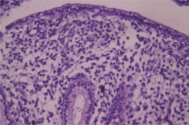
Histopathological examination of the 16 specimens taken in the proliferative phase showed the endometrial glands straight and narrow. They were lined by columnar cell with basal oval nuclei (Figure 1).
Meanwhile, in the 14 specimens taken during the secretory phase of the menstrual cycle, the endometrial glands appeared wider and showed some sacculations and cytoplasmic vacuolation. The nuclear cytoplasmic ratio of the cells lining the endometrial glands was 1:2.28 + 0.25.
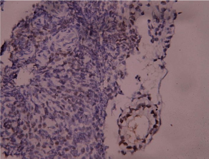
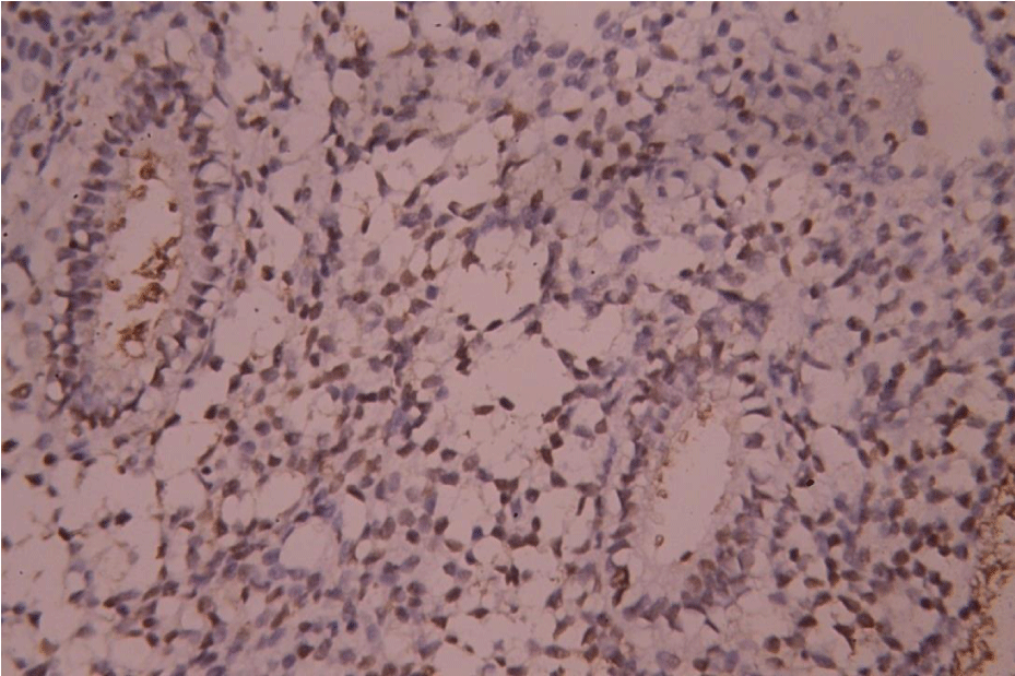
Immunohistochemical study of the hormone receptor expression in the controls was done. ER alpha receptor expression was seen in the nuclei of some of the cells in the endometrial glands and stroma that showed brown coloration. Also, the cells with positive progesterone receptor expression showed the hormone expression in the nuclei that appeared brown in color.
The percent of the positively stained nuclei expressing ER alpha and the intensity of its coloration is shown in table 1. The percent of the positively stained nuclei expressing PR and the intensity of its coloration is shown in table 2.
ER alpha expression (number of positive cells and intensity of reaction) is significantly higher in the endometrial glands as compared to stroma. However, PR expression (number of positive cells and intensity of reaction) is significantly higher in the endometrial stroma than the endometrial glands (Figure 2,3).
The females in the group of Abnormal Uterine Bleeding (AUB) group had a mean age of 32.42 years (range 25-40). Twelve of them were complaining of the 1st attack of abnormal uterine bleeding continuing for more than 8 days while 18 were complaining of recurrent 2nd or more attack of abnormal uterine bleeding. Twenty had prolonged or heavy menstrual bleeding while 10 had irregular bleeding.
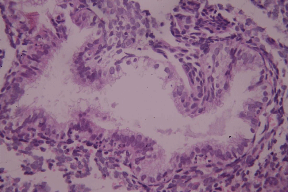
The shape of the endometrial glands in the different cases was examined. There was an apparent increase in the glandular content within the endometrium. The endometrial glands appeared wide relatively long and with irregular wall in 14 cases. In 5 cases, the endometrial glands appeared narrow and long while they appeared short and narrow in 2 cases. In 9 cases, the endometrial glands showed sacculations and cytoplasmic vacuolations (Figure 4). The mean nuclear /cytoplasmic ratio was 1:2.16 ± 0.22.
On comparing with the control group using independent student t test, there was a non-significant difference in the nuclear cytoplasmic ratio between females with dysfunctional uterine bleeding and females of the control group (t = 1.65, p = 0.10).
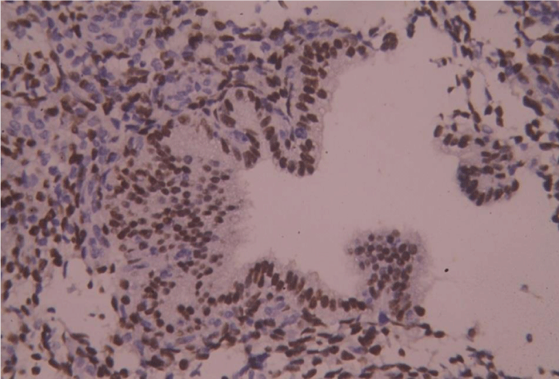
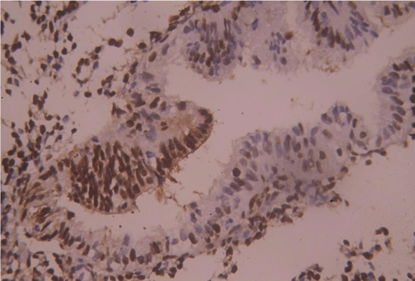
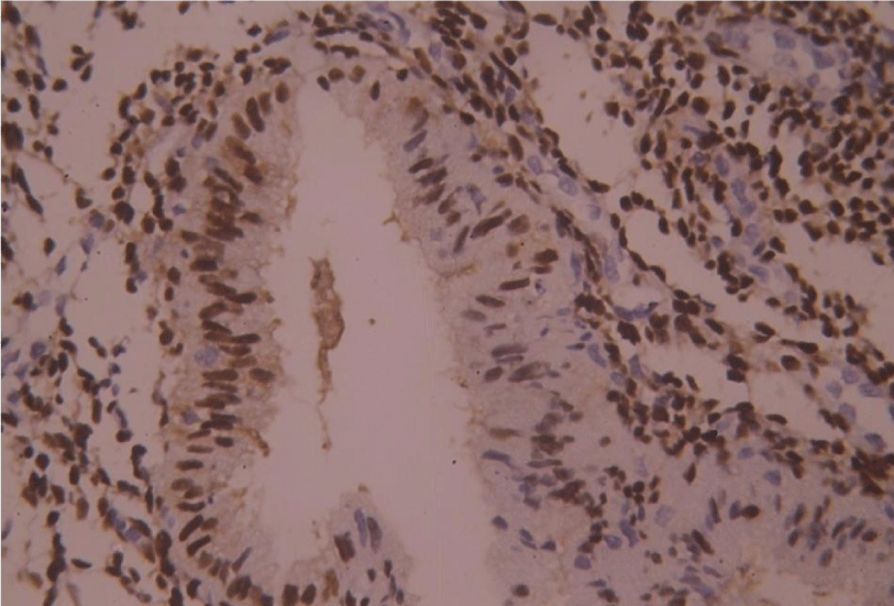
Immunohistochemical study was done for the estrogen receptor alpha and progesterone receptor expression. The results showed that they were expressed in the nuclei that appeared brown in color (Figure 5-7).
There was a significant increase in ER alpha expression in both endometrial glands and stroma in females with abnormal uterine bleeding as compared to control females.
There is a significant increase in PR expression in the endometrial glands and stroma in females with abnormal uterine bleeding as compared to the females in the control group.
Discussion
AUB is a common health problem in females that has a negative impact on their quality of life. Finding the cause of the AUB could help in trying medical treatment instead of surgery. In this study we evaluated the histopathological characters of the endometrium and its expression of ER alpha and PR receptors which could pave the way for appropriate medical treatment of these conditions.
Most of the females with AUB examined in our study showed wide relatively long glands with occasional sacculations in 30% of cases. Similar study reported cylindrical endometrial glands with simple endometrial hyperplasia in females with dysfunctional uterine bleeding [4]. However, in cases with endometrial carcinoma, the glands appeared extensively ramifying [4].
A study was done in Turkey on 307 females with abnormal uterine bleeding by Ozer et al. [5]. They found proliferative endometrium in 36.2% of cases, secretory endometrium in 27.4%, decidualization in 10.1%, endometrial polyps in 8.8%, endometritis in 6.5%, endometrial hyperplasia in 4.2%, irregular shedding in 3.9%, atrophic endometrium in 1.3%, placental retention in 1% and endometrial carcinoma in 0.7%.
The endometrium structure is affected by the different steroid hormones especially the estrogen and progesterone. There are 2 isoforms of estrogen receptors; ER alpha and ER beta. Matsuzaki et al. [6] underwent a quantitative analysis of the messenger ribonucleic acid of ER alpha and ER beta in the normal endometrium and in ovarian endometriotic cysts. They found that the normal endometrium contains significantly higher mRNA of ER alpha than mRNA of ER beta. Moreover, there are cyclic changes in their levels with the cyclic changes in ovarian hormones. Therefore, the principal regulatory effects of estrogens are mediated mainly by ER-alpha rather than ER-beta in both the eutopic endometrium and endometriotic cysts. Therefore, in this study we chose to examine the ER alpha isoform of estrogen receptors.
Estrogen and progesterone hormone receptors are involved in the autocrine and paracrine control of endometrial glands in response to estrogen and progesterone hormones [7]. This indicates that these receptors and estrogen and progesterone hormone play the important role in endometrial physiology. There are cyclic variations during the menstrual cycle as there is a significant decrease in ER alpha and PR expressions from the proliferative to the late secretory phase [7].
Regarding progesterone receptor expression, Godinjak and Bilalovic et al. [8] found that there are changes in the endometrial glands while they remain relatively constant in the endometrial stroma indicating difference in sensitivity of glandular from stromal progesterone receptors to hormone regulation during the menstrual cycle.
Generally, we found that the ER alpha expression was significantly higher in the endometrial glands than in the endometrial stroma. Meanwhile, the PR expression in the endometrial stroma was significantly higher than in the endometrial glands.
In our study, there was a significant increase ER alpha expression in the endometrial glands in patients with AUB as compared to the controls. In women with dysfunctional uterine bleeding with anovulation, there is continuous stimulation of the endometrial glands by estrogen hormone without the opposing effect of progesterone. This leads to proliferation of the endometrial glands in various degrees with repeated bleeding [9].
Unopposed estrogen hormone stimulatory effect results in Dysfunctional Uterine Bleeding (DUB). Patients with DUB had a significant higher endometrial thickness and ER and PR levels in the endometrium [10]. Moreover, there are higher ER and PR levels in hyperplastic glands. However, the receptor levels were low in hyperplastic glands showing atypia indicating that cases with atypical hyperplasia have a down-regulation of ER and PR receptors. This could indicate a precursor lesion to carcinoma and they do not respond to hormone therapy.
The increased estrogen receptor expression in females with dysfunctional uterine bleeding could be explained by that most of them have anovulatory cycle. In these cases there is no corpus luteum and therefore no progesterone hormone so the effect of estrogen is unopposed [4]. Without the progesterone hormone, the endometrial glands will proliferate and there will be outgrowing their blood supply. These glands will slough resulting in irregular bleeding that may be prolonged [11].
Women with various gynecologic disorders show abnormal uterine receptivity and abnormal endometrial biomarkers expression. Certain types of uterine receptivity defects were found to be caused by loss of ER alpha down regulation in the mid-secretory phase [12]. This could explain our findings in females with AUB who showed high ER alpha expression although the endometrial glands appeared wide and sacculated as that of secretory endometrium.
Treatment of AUB can include medical therapies and avoid surgical procedures [3]. Histopathology is the gold standard in diagnosis of the cause of abnormal uterine bleeding [13]. It is clear the immunohistochemical techniques are beneficial in assessing tissue localization, distribution and intensity of estrogen and progesterone hormone receptors in glandular and stromal cells of the endometrium. The examination of estrogen and progesterone receptor expression is more important than the biochemical examination of the hormone itself because it gives an idea about the actual effects of these hormones [14]. Therefore, it is a useful investigation on which clinical trials could be done to try medical treatment of AUB in reproductive age group.
Recommendation
Pelvic ultrasonography, histopathology and immuno-histochemical study of the receptors in endometrial biopsies are important tools for the evaluation of a case of abnormal uterine bleeding during the reproductive age group.
- Levy Zauberman Y, Pourcelot AG, Capmas P, Fernandez H. Update on the management of abnormal uterine bleeding. J Gynecol Obstet Hum Reprod. 2017; 46: 613-622. https://goo.gl/ZY2MUE
- Whitaker L, Critchley HO. Abnormal uterine bleeding. Best Pract Res Clin Obstet Gynaecol. 2016; 34: 54-65. https://goo.gl/zDo4tw
- Liu Z, Doan QV, Blumenthal P, Dubois RW. A systematic review evaluating health-related quality of life, work impairment, and health-care costs and utilization in abnormal uterine bleeding. Value Health. 2007; 10: 183-194. https://goo.gl/9hncqP
- Chakravarthy VK, Nag U, Rao DR, Anusha AM. Estrogen and progesterone receptors in dysfunctional uterine bleeding. Journal of Dental and Medical Sciences. 2013; 4: 73-76. https://goo.gl/zYA4vx
- Ozer A, Ozer S, Kanat Pektas M. Correlation between transvaginal ultrasound measured endometrial thickness and histopathological findings in Turkish women with abnormal uterine bleeding. J Obstet Gynaecol Res. 2016; 42: 573-578. https://goo.gl/46LXm8
- Matsuzaki S, Uehara S, Murakami T, Fujiwara J, Funato T, Okamura K. Quantitative analysis of estrogen receptor alpha and beta messenger ribonucleic acid levels in normal endometrium and ovarian endometriotic cysts using a real-time reverse transcription-polymerase chain reaction assay. Fertil Steril. 2000; 74: 753-759. https://goo.gl/3LQug9
- Mylonas I, Jeschke U, Shabani N, Kuhn C, Balle A, Kriegel S, et al. Immunohistochemical analysis of estrogen receptor alpha, estrogen receptor beta and progesterone receptor in normal human endometrium. Acta Histochem. 2004; 106: 245-252. https://goo.gl/8vmwfK
- Godinjak Z, Bilalovic N. Estrogen and progesterone receptors in endometrium in women with unexplained infertility. Mater Sociomed. 2014; 26: 51-52. https://goo.gl/2p9rmA
- Yin G, Zhu T, Li J, Chen M, Yang S, Zhao X. Decreased expression of survivin, estrogen and progesterone receptors in endometrial tissues after radiofrequency treatment of dysfunctional uterine bleeding. World J SurgOncol. 2012; 10: 100. https://goo.gl/ZAwaZ4
- Chakraborty S, Khurana N, Sharma JB, Chaturvedi KU. Endometrial hormone receptors in women with dysfunctional uterine bleeding. Arch Gynecol Obstet. 2005; 272: 17-22. https://goo.gl/6sMBHW
- Singh P, Singh P, Chaurasia A, Dhingra V, Misra V. Expression of ERα and PR in various morphological patterns of abnormal uterine bleeding-endometrial causes in reproductive age group. J Clin Diagn Res. 2016; 10: 6-9. https://goo.gl/cnMMu9
- Lessey BA, Palomino WA, Apparao KB, Young SL, Lininger RA. Estrogen receptor-alpha (ER-alpha) and defects in uterine receptivity in women. Reprod Biol Endocrinol. 2006; 4: 59. https://goo.gl/VXBb6B
- Khan R, Sherwani RK, Rana S, Hakim S, S Jairajpuri Z. Clincopathological patterns in women with dysfunctional uterine bleeding. Iran J Pathol. 2016; 11: 20-26. https://goo.gl/g1Hd9E
- Thakur B, Kaushik S, Garg S, Kishore S. Distribution pattern of ER & PR immunoexpression in endometrial biopsies of dub and infertile patients from a tertiary care centre. Annals of Pathothology and laboratory medicine. 2017; 4: 626-631. https://goo.gl/RsqzQG

Sign up for Article Alerts