Recurrent Vaginal Metastases after Cytoreductive Nephrectomy and Target Therapy with Sunitinib: Case Report?
Jesus Fernando Nagore Ancona1*, Jesus Antonio Martin Perez1, Rodrigo Moreno Garcia2, Denisse Anorve Bailon3, Diana Mendoza Elizarraraz4, Josue Andres Gonzalez Luna1, Ana Laura Sanchez Baltazar1 and Luisana Perna Lozada1
1Department of Surgery, Hospital Regional “General Ignacio Zaragoza” ISSSTE, CDMX, Mexico
2Department of Surgical Oncology, Hospital Regional “Gral Ignacio Zaragoza” ISSSTE, CDMX, Mexico
3Department of Medical Oncology, Hospital Regional “Gral Ignacio Zaragoza” ISSSTE, CDMX, Mexico
4Department of Pathology, Hospital Regional “Gral Ignacio Zaragoza” ISSSTE, CDMX, Mexico
*Address for Correspondence: Nagore Ancona, Jesus Fernando, Department of Surgery, Hospital Regional “General Ignacio Zaragoza” Social Services and Security Institute for the State Employees (ISSSTE), Mexico, Calz. Ignacio Zaragoza 1711, Ejercito Constitucionalista, Chinam Pac de Juarez, 09220, Mexico, ORCid: 0000-0002-7582-0172; Tel: + 556-198-2846; 558-805-1766; E-mail: [email protected]
Submitted: 03 September 2020; Approved: 10 September 2020; Published: 11 September 2020
Citation this article: Nagore Ancona JF, Martin Perez JA, Garcia RM, Bailon DA, Elizarraraz DM, et al. Recurrent Vaginal Metastases after Cytoreductive Nephrectomy and Target Therapy with Sunitinib: Case Report. Int J Case Rep Short Rev. 2020; 6(9): 056-00. https://dx.doi.org/10.37871/ijcrsr.id84
Copyright: © 2020 Nagore Ancona JF, et al. This is an open access article distributed under the Creative Commons Attribution License, which permits unrestricted use, distribution, and reproduction in any medium, provided the original work is properly cited
Keywords: Renal cell carcinoma; Secondary vaginal neoplasms; Immunohistochemistry
Download Fulltext PDF
Introduction: Renal cell carcinoma is a malignant neoplasm that originates from the renal tubular epithelium. Comprises approximately 2-3% of all adult cancers and has the highest mortality of all genitourinary cancers.
Methods: A 57-year-old, female, presents hematuria in August 2017. A CT Scan reveals right renal mass a right radical nephrectomy was performed. Initiates Sunitinib 50 mg per day, with partial response.
Results: In May 2019 presents vaginal bleeding, physical examination shows a vaginal mass, located in the lower third on the anterior wall, histopathological examination of metastasectomy reports poor differentiated clear cell carcinoma. In Abril 2020 recurrent bleeding was present, a new vaginal mass was seen, and totally resected with histopathological report of poor differentiated clear cell carcinoma. Immunohistochemistry was not performed in either case.
Conclusion: A clinical case of mRCC is described, developing clinical trials will provide new evidence in order to modify current guidelines in the management of kidney cancer.
Abbreviations
CT: Computed Tomography; mRCC: Metastatic Renal Cell Carcinoma
Introduction
Renal Cell Carcinoma (RCC) is a malignant neoplasm that originates from the renal tubular epithelium. The classic presentation of RCC is flank pain, hematuria, and a palpable abdominal mass. However, this presentation is uncommon and occurs in less than 10% of affected individuals [1]. Risk factors include End-Stage Renal Disease (ESRD), Acquired Cystic Kidney Disease (ACKD), smoking, hypertension, occupational exposure and genetic factors, among others. There are approximately 400,000 newly diagnosed cases of kidney cancer each year, with RCC accounting for the vast majority of cases. RCC comprises approximately 2-3% of all adult cancers and has the highest mortality of all genitourinary cancers. It has an indolent course in many patients, one third of patients already have advanced disease that is locally invasive or metastatic at presentation, and remains an important cause of cancer-related death with a 5-year survival rate of approximately 8% [2,3].
The most common metastatic sites are described in table 1. Among the unusual sites of metastasis, the vagina is a rare localization; only a little number of cases has been reported in the literature. The tumoral bleeding is a symptom present in 6 to 10% of patients. Such hemorrhagic events may decrease quality of life, require hospitalization, and in extreme cases lead to death. The hemostatic radiotherapy has appeared relevant as a therapeutic strategy in palliative supportive care, being an indication in case of tumoral bleeding [4].
It is postulated that retrograde venous flow via the ovarian vein facilitates seeding of the vaginal mucosa from the kidneys, making left sided RCC more likely to result in vaginal metastasis due to aberrant flow between the left ovarian vein and the left renal vein. Radiological studies have observed flux in venous flow in patients with RCC accompanied by vaginal metastasis. The individual under consideration presented with vaginal cancer in the setting of left-sided RCC, further adding to the legitimacy of this unique mechanism of spread. Regardless of etiology, vaginal cancer presents with bleeding that can be minimal to profuse [5].
In order to provide the best treatment for patients with metastasic RCC based on the current literature, we discuss the management employed a 57 years old woman with vaginal metastasis 2 years after cytoreductive nephrectomy follow by target therapy.
Case Presentation
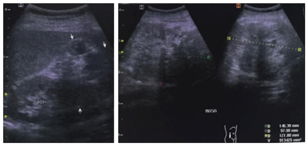
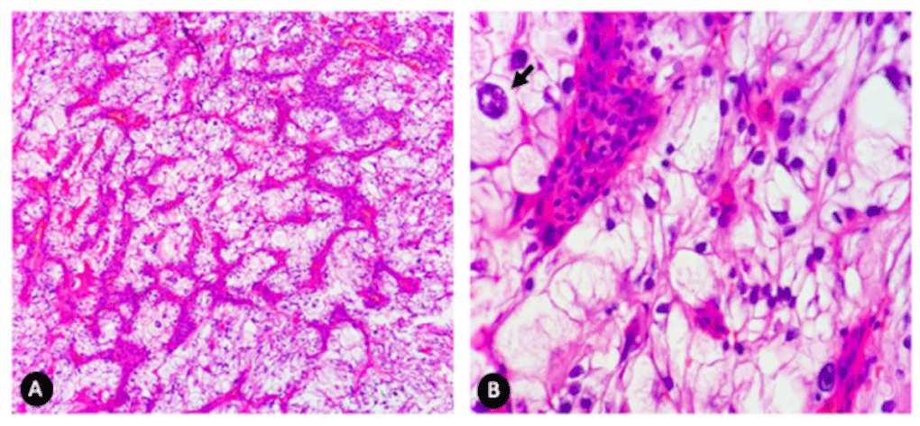
A 57-year-old, female, non-current smoker (2 cigarettes daily for fourty two years), presents hematuria in August 2017. Past medical history was unremarkable. An ultrasound shows a suspicious ectasia in the right kidney and no lithiasis (Figure 1), therefore requesting a CT Scan which reveals right renal mass, heterogeneuos, aumented vascularity, changes in the perirrenal fat and involvent of the right renal vein. Multiple pulmonary nodules, measuring up to 10 mm, were observed on imaging. A right radical nephrectomy was performed in November 2017. The tumor was attacheted to renal hilius, inferior vena cava, liver, psoas muscle, and right colon. Pathologic examination confirmed the Clear Cell Renal Carcinoma (ccRCC), Furhman grade was 2. The tumor invaded into the perinephric fat tissue, measuring 13.0 x 11.0 x 8.0 cm. A diagnosis of metastatic ccRCC was made. The final pathological stage was pT3a pNx pM1. In January 2018 inities first-line treatment, Sunitinib 50 mg per day, four treatment weeks and two rest weeks, titrating the dose due to side effects (Grade 2 Mucositis). Pulmonary nodes surveillance with CT Scan and PET/CT with full radiologic response. In May 2019 presents vaginal bleeding, physical examination shows a vaginal mass, located in the lower third on the anterior wall, measuring approximately 2.0 x 2.0 cm in size. Biopsy was performed with histopathology. Metastases of clear cell carcinoma (Figure 2). She was referred for metastasectomy, performed in July 2019. On gross examination tumor of 1.0 x 1.0 cm, borders up to 0.2 cm. PET/CT in November 2019 without any evidence that suggest infiltrative malignancy. In April 2020 recurrent bleeding was present, a new vaginal mass was seen, and totally resected with histopathological report of poor differentiated clear cell carcinoma. Immunohistochemistry was not performed in either case. There was no data of progression in other sites, so it was decided to treat with vaginal brachitherapy, which has not been started to the present day.
Discussion
The National Comprehensive Cancer Network (NCCN) recommends individualizing cytoreductive nephrectomy based on symptoms and extent of metastatic disease. Generally, patients who would be candidates for Cytoreductive Nephrectomy (CN) prior to systemic therapy per NCCN are patients with excellent performance status and no brain metastasis. Patients with metastatic disease who present with hematuria or a symptomatic primary tumor should be offered palliative nephrectomy if they are surgical candidates. In regards to non-clear cell RCC, the NCCN, and European Association for Urology (EAU) make no specific recommendations for CN [5].
Two aleatorized trials both published in 2001 by Flanigan et al and Mickisch et al. established the benefit of cytoreductive nephrectomy for patients with metastatic Renal Cell Carcinoma (mRCC) however, these patients were treated with interferon a-2b [6,7]. More recently, the CARMENA trial, a randomized phase 3 trial, which did not show a survival benefit when cytoreductive nephrectomy was performed prior to treatment with sunitinib (Overall survival CN + Sunitinib 15.6 months vs 19.8 months in the only Sunitinib arm). The findings of this study, which included patients with intermediate–poor-risk disease, emphasize the importance of patient selection for cytoreductive nephrectomy [8]. Table 2 describes the Stage-based survival percentage by years. Despite the heterogenicity of evaluated studies, ultimately, several genetic mutations have been identified as being associated with differential clinical outcomes with different targeted therapies and immunotherapies. Sunitinib malate, an oral, multitargeted receptor Tyrosine Kinase Inhibitor (TKI), has been the gold-standard first-line treatment for mRCC for the past 12 years [2,9].
The renal cell carcinoma has the capability to metastasize throughout the body, sometimes without preexisting knowledge of the primary tumor. Although the morphologic appearance of classic clear-cell renal cell carcinoma (ccRCC) is distinctive in the kidney, histologic recognition of that entity can sometimes be difficult elsewhere [10]. The histologic differences between primary vaginal clear cell carcinomas and metastatic ccRCC vaginal lesions are subtle and differentiation in a small biopsy can be challenging. Typically vaginal Clear Cell Carcinoma (CCC) show variable morphologic patterns including solid, tubulocystic, and papillary, with presence of hobnail cells. ccRCC characteristically shows alveolar, acinar, and nested patterns and papillary architecture is not a feature of ccRCC in addition, a prominent network of branching small, thin-walled blood vessels is characteristic and diagnostically helpful. Therefore, due to the presence of extensive overlapping morphologic features between both tumors, it is recommended to have the kidneys evaluated in cases of vaginal neoplasms with clear cell features. This examination would thus prevent a misdiagnosis of a primary vaginal adenocarcinoma. Immunohistochemistry is helpful in differentiating these entities as CA-IX and CD10 are most frequently expressed in ccRCC than Mullerian CCC, whereas CK7, Napsin-A, and methylacyl-coenzyme-A racemase (AMACR) show a reverse pattern of expression. Although PAX8 is a very important marker for the diagnosis of mRCC, it is also expressed in CCC of Mullerian origin and therefore is of no use in distinguishing a renal versus a vaginal primary [1,3].
Immunohistochemistry stands out, complementary to histopathological diagnosis by morphology only, since due to a common embryological origin, is not entirely possible to determinate a metastatic nature of the lesion or a second primary, being antibodies reported by multiple authors, therefore immunohistochemistry being mandatory. A second primary, CCC of the vagina, should be treated as followed: The type of surgical therapy is also chosen based on the site and range of occurrence of the primary lesion. At this time the physician must consider both removal of the primary lesion and regional lymph nodes simultaneously. In particular, in the case of vaginal cancer occurring in the upper third of the vagina, surgical therapy consisting of hysterectomy extended to the vagina is a good option, nevertheless in this case, lesion was 2 cm up from the introitus, so hysterecomy and vaginectomy was not indicated, nethier pelvic exanteration.
The surveillance using PET/CT in the current work does not detect the vaginal mass as hypermetabolic activity. The vaginal bleeding and foreign body sensation, described by the patient, guides the clinical diagnosis. The necesity of PET/CT remains in doubt, whether conventional CT scan can be sufficient as a follow-up imaging tool. Colposcopy is not indicated for follow-up, unless specific symptoms such as pain or vaginal bleeding are present.
In the literature, possible hematogenous dissemination pathways of RCC to the vagina are described, ruling out that exact mechanism is still unknown [11]. In consideration of the authors, these dissemination routes consist only as historical background, since currente advances in genomics describe gene mutations in the appearence of renal cell carcinoma in distant organs as well, according to Bracarda S, et al. [12].
In regards of the reviewed literature, Rehailia-Blanchard, et al. described the employement of radiotherapy in their case report as hemostatic therapy, and in addition to Sunitinib evidenced a 40% decrease of the vaginal metastasis, with a concomitant bleeding stop. Tselis and Chatzikonstantinou in their analysis highlighted the substantial limitations of the various studies including the use of non-conformal RT techniques, inappropriate dosing, and outdated technology, concluding the need for multiinstitutional trials to investigate the additional benefits of adjuvant RT regarding overall survive along with targeted therapy, concluding the addition of RT to immunotherapy may potentiate the generation of antitumor immune responses, which could treat existing metastases as well as prevent future metastases [13].
Conclusion
A clinical case of RCC is described, with pulmonary nodules, therefore, metastatic at the time of the diagnosis, despite cytoreductive nephrectomy and target therapy, subsequent vaginal metastasis appear, completely resected. Developing clinical trials will provide new evidence in order to modify current guidelines in the management of kidney cancer.
- Jimenez AR, Rivera Rolon MDM, Eyzaguirre E, Clement C. Vaginal bleeding as initial presentation of an aggressive renal cell carcinoma: A case report and review of the literature. Case rep pathol. 2018: 2109279. DOI: 10.1155/2018/2109279
- Moran M, Nickens D, Adcock K, Bennetts M, Desscan A, Charnley N, et al. Sunitinib for metastatic renal cell carcinoma: A systematic review and meta-analysis of real-world and clinical trials data. Target Oncol. 2019; 14: 405-416. DOI: 10.1007/s11523-019-00653-5
- Machiele R, Renbarger T, Guidry B. Severe vaginal bleeding in a case of renal cell carcinoma. Case Rep Obstet Gynecol. 2019: 2174051. DOI: 10.1155/2019/2174051
- Rehailia-Blanchard A, Morel A, Rancoul C, He MY, Magne N, Falkowski S. Vaginal metastasis of renal clear-cell cancer. Gulf J Oncolog. 2018; 1: 67-71. PubMed: https://pubmed.ncbi.nlm.nih.gov/29607827/
- Alhalabi O, Karam JA, Tannir NM. Evolving role of cytoreductive nephrectomy in metastatic renal cell carcinoma of variant histology. Curr Opin Urol. 2019; 29: 521-525. DOI: 10.1097/MOU.0000000000000661
- Flanigan RC, Salmon SE, Blumenstein BA, Bearman SI, Roy V, McGrath PC, et al. Nephrectomy followed by interferon alfa-2b compared with interferon alfa-2b alone for metastatic renal-cell cancer. N Engl J Med. 2001; 345: 1655-1659. DOI: 10.1056/NEJMoa003013
- Mickisch GH, Garin A, van Poppel H, de Prijck L, Sylvester R. Radical nephrectomy plus interferon-alfa-based immunotherapy compared with interferon alfa alone in metastatic renal-cell carcinoma: A randomised trial. Lancet. 2001; 22; 358: 966-970. DOI: 10.1016/s0140-6736(01)06103-7
- Mejean A, Ravaud A, Thezenas S, Colas S, Beauval JB, Bensalah K, et al. Sunitinib alone or after nephrectomy in metastatic renal-cell carcinoma. N Engl J Med. 2018; 379: 417-427. DOI: 10.1056/NEJMoa1803675
- Mano R, Gopal N, Hakimi AA. The evolving role of cytoreductive nephrectomy: Incorporating genomics of metastatic renal cell carcinoma into treatment decisions. Curr Opin Urol. 2019; 29: 531-539. DOI: 10.1097/MOU.0000000000000663
- Mentrikoski MJ, Wendroth SM, Wick MR. Immunohistochemical distinction of renal cell carcinoma from other carcinomas with clear-cell histomorphology: Utility of CD10 and CA-125 in addition to PAX-2, PAX-8, RCCma, and adipophilin. Appl Immunohistochem Mol Morphol. 2014; 22: 635-641. DOI: 10.1097/PAI.0000000000000004
- Martinez Ruiz J, Ruiz Mondejar R, Carrion Lopez P, Martinez Sanchiz C, Donate Moreno MJ, Pastor Navarro H, et al. Metastasis in the vagina of renal tumor of cellulas claras. Arch Esp Urol. 2011; 64: 380-383.
- Bracarda S, Porta C, Sabbatini R, Rivoltini L. Angiogenic and immunological pathways in metastatic renal cell carcinoma: A counteracting paradigm or two faces of the same medal? The GIANUS Review. Crit Rev Oncol Hematol. 2019; 139:149-157. DOI: 10.1016/j.critrevonc
- Tselis N, Chatzikonstantinou G. Treating the Chameleon: Radiotherapy in the management of Renal Cell Cancer. Clin Transl Radiat Oncol. 2019; 16: 7-14. DOI: 10.1016/j.ctro.2019.01.007
- Bianchi M, Sun M, Jeldres C, Shariat SF, Trinh QD, Briganti A, et al. Distribution of metastatic sites in renal cell carcinoma: A population-based analysis. Ann Oncol. 2012; 23: 973-80. DOI: 10.1093/annonc/mdr362
- Vidart A, Fehri K, Pfister C. Unusual metastasis of renal carcinoma. Ann Urol (Paris). 2006; 40: 211-219. DOI: 10.1016/j.anuro.2006.03.004


Sign up for Article Alerts