Can we Build a House From one Brick ? : Diagnosis of TGV Diagnosis from a Single Stored Heart Volume
Wael El Guindi1, Sameh Elkhot2, Michel Dreyfus3, Sondos Salem4, Amr Abasy5 and Mahmoud Alalfy6
1Consultant in Obstetrics and Gynecology, Franck Joly Hospital, France
2Lecturer of Obstetrics and Gynecology, Cairo University Hospital, Egypt
3Professor of Obstetrics and Gynecology, Caen University Hospital, Caen, France
4Lecturer of Obstetrics and Gynecology, National Research Centre, Egypt
5Assistant professor of Obstetrics and Gynecology, National Research Centre, Egypt
6Head of the Fetal medicine unit, National Research Centre, Egypt
*Address for Correspondence: Wael El Guindi, Department of Obstetrics and Gynecology, Franck Joly Hospital, Saint-Laurent-du-Maroni, France, Paris, Tel. No: 0774253858; E-mail: [email protected]
Submitted: 06 December 2020; Approved: 24 December 2020; Published: 28 December 2020
Citation this article: El Guindi W, Elkhot S, Dreyfus M, Salem S, Abasy A, Alalfy M. Can we Build a House From one Brick ? : Diagnosis of TGV Diagnosis from a Single Stored Heart Volume. Int J Cardiovasc Dis Diagn. 2020 Dec 28;5(2): 047-053.
Copyright: © 2020 Guindi W, et al. This is an open access article distributed under the Creative Commons Attribution License, which permits unrestricted use, distribution, and reproduction in any medium, provided the original work is properly cited
Disclaimer: The opinions and assertions expressed herein are those of the authors and do not necessarily reflect the official policy or position of the Uniformed Services University or the Department of Defense.
Download Fulltext PDF
We present a case in which D-TGV was diagnosed by off-line analysis from only one stored 3D volume. To our knowledge, this is the first study to reconstruct the cardiovascular anatomy and establish the diagnosis of complex cardiac malformation by offline analysis of only one stored 3 D volume.
Introduction
The dextro-transposition of the great vessels is a structural heart defect with an atrio-ventricular concordance and ventriculo-arterial discordance, it is the most common type of TGV, the aorta arises from the right ventricle and the pulmonary artery arises from the left ventricle. It the second most common neonatal cyanotic congenital heart disease representing 5–7% of all CHDs [1]. D-TGV is a critical heart defect lesion that should be diagnosed prenatally, as this allows for scheduling of delivery in a specialist center with with the possibility of offering an urgent Balloon Atrial Septostomy (BAS) if necessary. The sequelae of TVG could be severe if undiagnosed prenatally with mortality approaching 85% to 90%a (85-90%) if not treated [2].
Despite the progress in prenatal diagnosis, antenatal detection rate of TGA/IVS has improved but still remains less than 50% of patients [3].
Case
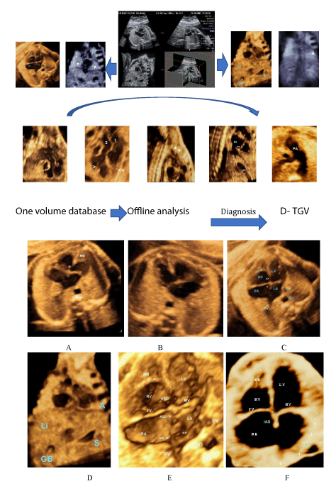
A pregnant woman of 22 weeks gestation arrived in emergency department for abdominal pain. During the initial ultrasound examination performed by an experienced sonographer who was in charge, interventricular communication was observed, volume data is stored, and the sonographer had to discontinue the examination due to an emergency cesarean section. The patient did not show up for her appointment at the prenatal diagnostic unit and could not be reached after several attempts. We have tried to gather as much information as possible from the single stored volume of the 4-chamber view. Offline analysis of the only volume database that we had showed situs solitus, levocardia with a normal cardiac axis. On performing offline analysis, the four-chamber view revealed atrioventricular concordance, normal atrioventricular valves, and well-sized ventricles. The Pulmonary Artery (PA) originating from the left ventricle, the aorta of the right ventricle and the aorta was to the left of the PA confirming the presence of TGA. Ultrasound images were then sent via the Internet to a reference center for prenatal diagnosis (CHU de Caen, France), and the initial diagnosis was confirmed by a multidisciplinary team comprising a pediatric cardiologist, and a pediatric cardiac surgeon. The patient could have finally been reached and she was followed in our antenatal unit and subsequent ultrasound examinations confirmed the diagnosis. The family was informed in detail of the postnatal corrective surgical treatment available. Amniocentesis was performed and revealed a normal male karyotype. The patient was sent to deliver in a university teaching hospital in Paris. She gave birth to a full-term male baby weighing 3.1 kg. and the diagnosis of TGV was confirmed. At birth, prostaglandin E1 was administered initially but was discontinued after the septostomy. Neonatal arterial switch operation (ASO) was performed. The operative consequences were simple. He was followed until the age of 7, he was in good health.
Discussion
The introduction of 3- and 4-dimensional (3D / 4D) ultrasound, in particular Spatial And Temporal Image Correlation (STIC) about 15 years ago [4], has added several advantages to fetal echocardiography, in particular the ability to extract a large number of ultrasound plans from 3D / 4D volumes. The integration of 3D / 4D into fetal echocardiography has been used to display images in surface mode with two-dimensional (2D) and color Doppler [5], to extract one or more planes for tomographic view [6], thus allowing the offline examination of the heart, remote diagnosis and facilitating scientific cooperation between high and low-income countries [7]. The medical and surgical management of TGA is well-established in the developing in the developing world is associated with an early survival of 85% [8]. On the other hand, lack of detection may increase morbidity and mortality rate [9]. The diagnosis of fetal TGA is closely related to operator skill and expertise and necessitates thorough knowledge of cardiac anatomy to allow accurate diagnosis and optimal management [10].
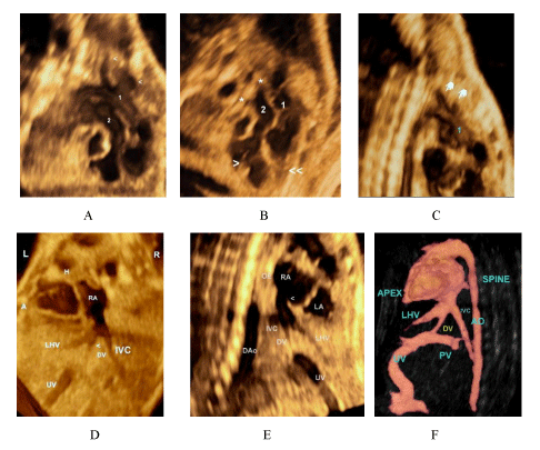
The cornerstone of diagnosis of TGV is based on the assessment of ventricular outflow tract and great vessels [11]. By definition, TGV means discordant connections between the ventricles and the great arteries, consequently, to establish a diagnosis it is necessary to highlight the anatomic features of both ventricles and great vessels. From the only volume stored we followed the segmental approach described in the literature [10]. We have reconstructed four-chamber views in such a way that the tip of the heart is oriented either to the left, levocardia, to the right, dextrocardia, i.e. indeterminate situs, (figure 1 A,B) then we were able to reconstruct the 3-dimensional anatomy in a sagittal section clearly showing a situs solitus with the point of the heart facing the left side, gall bladder on the right, stomach on the left and the liver is right-sided, i.e Situs Solitus (figure 1 C,D). From the stored volume we were able to demonstrate right ventricular moderator band , fibromuscular structures that traverse the cavity of the right ventricles (figure 1 C,E, figure 2B), left atrial appendage that has a broad-based triangular appearance (figure 1 C,E); the supra diaphragmatic part of IVC connecting the RA by DV (figure 2D), in addition to Eustachian Valve (EV) an embryonic structure redirecting the blood flow from the inferior vena cava through the foramen oval. (figure 2E).
We identified the left atrial appendage which is characteristically a slender finger-like with the presence of foramen oval flap (red arrow), and pulmonary veins draining into LA (figure 2 C,E).
Ventricular Outflow Tract Reconstruction showed parallel great vessels with loss of the normal crossover, vessel 1 originating from the right, continues upwards forming an arch that gives off branches then passes downward (descending aorta) towards the abdominal cavity, along the spine. The other vessel (2) arises from the left ventricle recognized by the anterior right muscle, gives a bifurcation, i.e. the pulmonary artery (figure 2 A,B,C). Four chamber view have shown a ventricular septal defect VSD, atrial septal defect ADS, in addition, we have demonstrated the more apical position of the tricuspid valve (figure1 E).
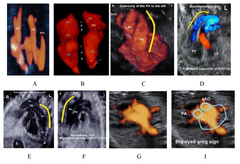
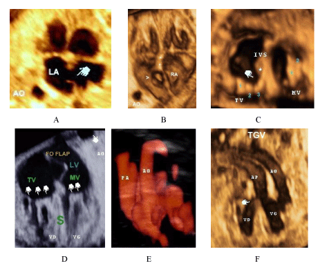
From a single stored volume we were able to establish the diagnosis of d-TGV, which was confirmed by subsequent ultrasound examinations, , In this context we would like to emphasize the importance of properly differentiating between d-TGV which requires immediate postnatal care in a specialized center and corrected L-TGA , in which there is an atrioventricular discordance and does not require immediate intervention as it does show symptoms until late in life until later in life [12]. From the subsequent ultrasound examinations of this patient we were able to confirm the diagnosis, in addition ,displaying cardiovascular anatomy using Power Doppler 3D Modes, moreover comparing images with standard ultrasound images of our patients serving as control cases to further clarify the diagnostic usefulness of 3DUS and doppler angiography and help to improve its antennal diagnosis, which remains modest despite the development in this domain , as half of cases of TVG are still not diagnosed antenatal (figure 1F,2F,3DE,4BD,5B).
From subsequent ultrasound studies of the same patient we demonstrated three signs
1- ”boomerang sign”: an abnormal right convexity of the aorta arising from the RV instead of the normal convexity to the left of the pulmonary artery arising from the RV observed in normal hearts [13] which is considered a reliable clue for diagnosing TGA (figure 4 B, C, D, F).
2- “baby bird’s beak image” described by McGahan JP et al, the pulmonary artery arises from the LV, the left branch makes a sharp angle with the main pulmonary artery and ductus arteriosus with the image of its bifurcation resembling the head of a baby bird with an open beak [14] (figure 3).
3- “big-eyed frog”, the great vessels were disposed side by side, resembling a Japanese fictional character, created by Sanrio, called “Keroppi” [15,16] (figure 4 G, I).
4. At last, we would like to point out that all the images in this study were carried out with a Voluson 730 Pro (General Electric, Milwaukee, WI, USA) with a volumetric abdominal transducer (4–8 MHz)., which does not contain the STIC software, i.e., without having access to STIC confirming the inherent capabilities of 3DUS, in addition we high lightened the importance of using this means to send a stored volume by internet to experts for remote diagnosis.
Conclusion
To our knowledge, this is the first study to reconstruct the cardiovascular anatomy and establish the diagnosis of complex cardiac malformation by offline analysis of only one stored 3 D volume. 3D ultrasound offers a high-resolution volume rendering image that provides excellent delineation of cardiac anatomy and add significantly to detection and understanding of the Cardiovascular anomalies. Offline analysis of cardiovascular anomalies conferred significant diagnostic advantages over standard 2D and represent an invaluable tool for the prenatal diagnosis and optimal management of fetuses with congenital heart diseases. This technology enabling worldwide remote diagnosis especially underserved area not having access to facilities allowing such a diagnosis and enhance scientific cooperation between high and low-income countries.
- Komarlu R., Morell V.O., Kreutzer J., Munoz R.A. (2020) Dextro-Transposition of the Great Arteries (D-TGA). In: Munoz R., Morell V., da Cruz E., Vetterly C., da Silva J. (eds) Critical Care of Children with Heart Disease. Springer, Cham. https://doi.org/10.1007/978-3-030-21870-6_32
- Komarlu R., Morell V.O., Kreutzer J., Munoz R.A. (2020) Dextro-Transposition of the Great Arteries (D-TGA). In: Munoz R., Morell V., da Cruz E., Vetterly C., da Silva J. (eds) Critical Care of Children with Heart Disease. Springer, Cham. https://doi.org/10.1007/978-3-030-21870-6_32
- Bravo-Valenzuela NJ, Peixoto AB, Araujo Júnior E. Prenatal diagnosis of transposition of the great arteries: an updated review. Ultrasonography. 2020 Oct;39(4):331-339. doi: 10.14366/usg.20055. Epub 2020 Jun 8. PMID: 32660209; PMCID: PMC7515665.
- DeVore GR, Falkensammer P, Sklansky MS, Platt LD. Spatio-temporal image correlation (STIC): new technology for evaluation of the fetal heart. Ultrasound Obstet Gynecol. 2003 Oct;22(4):380-7. doi: 10.1002/uog.217. PMID: 14528474.
- Chaoui R, Abuhamad A, Martins J, Heling KS. Recent Development in Three and Four Dimension Fetal Echocardiography. Fetal Diagn Ther. 2020;47(5):345-353. doi: 10.1159/000500454. Epub 2019 Jul 2. PMID: 31266014.
- Chaoui R, Heling KS. 3D-ultrasound in prenatal diagnosis: a practical approach. 1st edition. Berlin, New York: DeGruyter; 2016. https://www.degruyter.com/view/title/523221
- El Guindi W, Dreyfus M, Carles G, Lambert V, Herlicoviez M, Benoist G. 3D ultrasound and Doppler angiography for evaluation of fetal cardiovascular anomalies. Int J Gynaecol Obstet. 2013 Feb;120(2):173-7. doi: 10.1016/j.ijgo.2012.08.015. Epub 2012 Nov 11. PMID: 23151371.
- Schidlow DN, Jenkins KJ, Gauvreau K, Croti UA, Giang DTC, Konda RK, Novick WM, Sandoval NF, Castañeda A. Transposition of the Great Arteries in the Developing World: Surgery and Outcomes. J Am Coll Cardiol. 2017 Jan 3;69(1):43-51. doi: 10.1016/j.jacc.2016.10.051. PMID: 28057249; PMCID: PMC7144419.
- Escobar-Diaz MC, Freud LR, Bueno A, Brown DW, Friedman KG, Schidlow D, Emani S, Del Nido PJ, Tworetzky W. Prenatal diagnosis of transposition of the great arteries over a 20-year period: improved but imperfect. Ultrasound Obstet Gynecol. 2015 Jun;45(6):678-82. doi: 10.1002/uog.14751. Epub 2015 Apr 30. PMID: 25484180; PMCID: PMC4452393.
- Lapierre C, Déry J, Guérin R, Viremouneix L, Dubois J, Garel L. Segmental approach to imaging of congenital heart disease. Radiographics. 2010 Mar;30(2):397-411. doi: 10.1148/rg.302095112. PMID: 20228325.
- Ruxandra G, Toba M, Iliescu, Petru B. Anatomical consideration of the number and form of the papillary muscle in the left ventricle. ARS Medica Tomitana. 2016;22:119-127. https://content.sciendo.com/view/journals/arsm/22/2/article-p119.xml?language=en
- Szymanski MW, Moore SM, Kritzmire SM, et al. Transposition Of The Great Arteries. [Updated 2020 Aug 11]. In: StatPearls [Internet]. Treasure Island (FL): StatPearls Publishing; 2020. https://www.ncbi.nlm.nih.gov/books/NBK538434/
- Menahem S, Rotstein A, Meagher S. Rightward convexity of the great vessel arising from the anterior ventricle: a novel fetal marker for transposition of the great arteries. Ultrasound Obstet Gynecol. 2013 Feb;41(2):168-71. doi: 10.1002/uog.11171. PMID: 22492362.
- McGahan JP, Moon-Grady AJ, Pahwa A, Towner D, Rhee-Morris L, Gerscovich EO, Fogata M. Potential pitfalls and methods of improving in utero diagnosis of transposition of the great arteries, including the baby bird's beak image. J Ultrasound Med. 2007 Nov;26(11):1499-510; quiz 1511. doi: 10.7863/jum.2007.26.11.1499. PMID: 17957044.
- Shih JC, Shyu MK, Su YN, Chiang YC, Lin CH, Lee CN. 'Big-eyed frog' sign on spatiotemporal image correlation (STIC) in the antenatal diagnosis of transposition of the great arteries. Ultrasound Obstet Gynecol. 2008 Nov;32(6):762-8. doi: 10.1002/uog.5369. PMID: 18780310.
- Bravo-Valenzuela NJ, Peixoto AB, Araujo Júnior E. Prenatal diagnosis of transposition of the great arteries: an updated review. Ultrasonography. 2020 Oct;39(4):331-339. doi: 10.14366/usg.20055. Epub 2020 Jun 8. PMID: 32660209; PMCID: PMC7515665.


Sign up for Article Alerts