Safety with Green Zones in Hospital Medicine Practice Post Covid-19: A Case Series Report
Cara Hanley1, Seshnag Siddavaram2, Maha Khalid3 and Syed Husain4*
1Core Medical Trainee, Maidstone and Tunbridge Wells NHS Trust, UK
2Specialist Trainee Registrar, Maidstone and Tunbridge Wells NHS Trust, UK
3Specialty Doctor, Maidstone and Tunbridge Wells NHS Trust, UK
4Consultant Respiratory Physician, Maidstone and Tunbridge Wells NHS Trust, UK
*Address for Correspondence: Syed Husain, Consultant Respiratory Physician, Maidstone and Tunbridge Wells NHS Trust, Hon. Senior Clinical Lecture, Kings College London, Travers Unit, Maidstone Hospital, Hermitage Lane, Maidstone ME169QQ, Tel: +44-795-848-4422; Ext: +44-162-224-2591; ORCID ID: orcid.org/0000-0002-9953-9123; E-mail: [email protected]
Submitted: 21 July 2020; Approved: 11 August 2020; Published: 14 August 2020
Citation this article: Hanley C, Siddavaram S, Khalid M, Husain S. Safety with Green Zones in Hospital Medicine Practice Post Covid-19: A Case Series Report. Int J Cardiovasc Dis Diagn. 2020;5(1): 013-017.
Copyright: © 2020 Hanley C, et al. This is an open access article distributed under the Creative Commons Attribution License, which permits unrestricted use, distribution, and reproduction in any medium, provided the original work is properly cited
Download Fulltext PDF
We describe four complex cases of patients who had presented to hospital with a diagnostic dilemma wherein they came with signs and symptoms suggestive of COVID-19 infection with clinical, biochemical and radiological evidence of the infection but they had recurrent negative tests with nasopharyngeal RT-PCR swabs. Differential diagnoses were entertained in all patients and COVID-19 was not the only possibility in this group. We however feel that patients like these pose a challenge especially in the respiratory wards with aerosol generating procedures and a vulnerable patient cohort. Hence early identification and isolation of the suspected COVID-19 infections should take priority to ensure appropriate patient care is being delivered.
Background
The novel coronavirus SARS-CoV2 has caused significant global morbidity and mortality. The clinical course of the virus can range from a mild respiratory tract illness to full-blown acute respiratory distress syndrome (ARDS) in severe cases. Currently, the infection is confirmed using RT-PCR techniques which have shown a 30% false negative [1].
We describe four cases where standard oxygen therapy & Continuous Positive Airway Pressure (CPAP) was used in the treatment similar to COVID-19 patients despite having serial negative swabs to highlight the importance of looking at a holistic picture to make a diagnosis rather than relying on negative RT-PCR swabs alone to rule out the infection. This we feel will lead to appropriate clinical care being delivered when infection control measures are being followed.
Case Description
Case 1
A 71-year-old female non-smoker with no co-morbidities was admitted with of shortness of breath, pyrexia and a non-productive cough. She had been reviewed in the community and had completed a course of oral Amoxicillin. She developed tachycardia, tachypnoea and hypoxia with a type one respiratory failure ultimately requiring hospital admission.
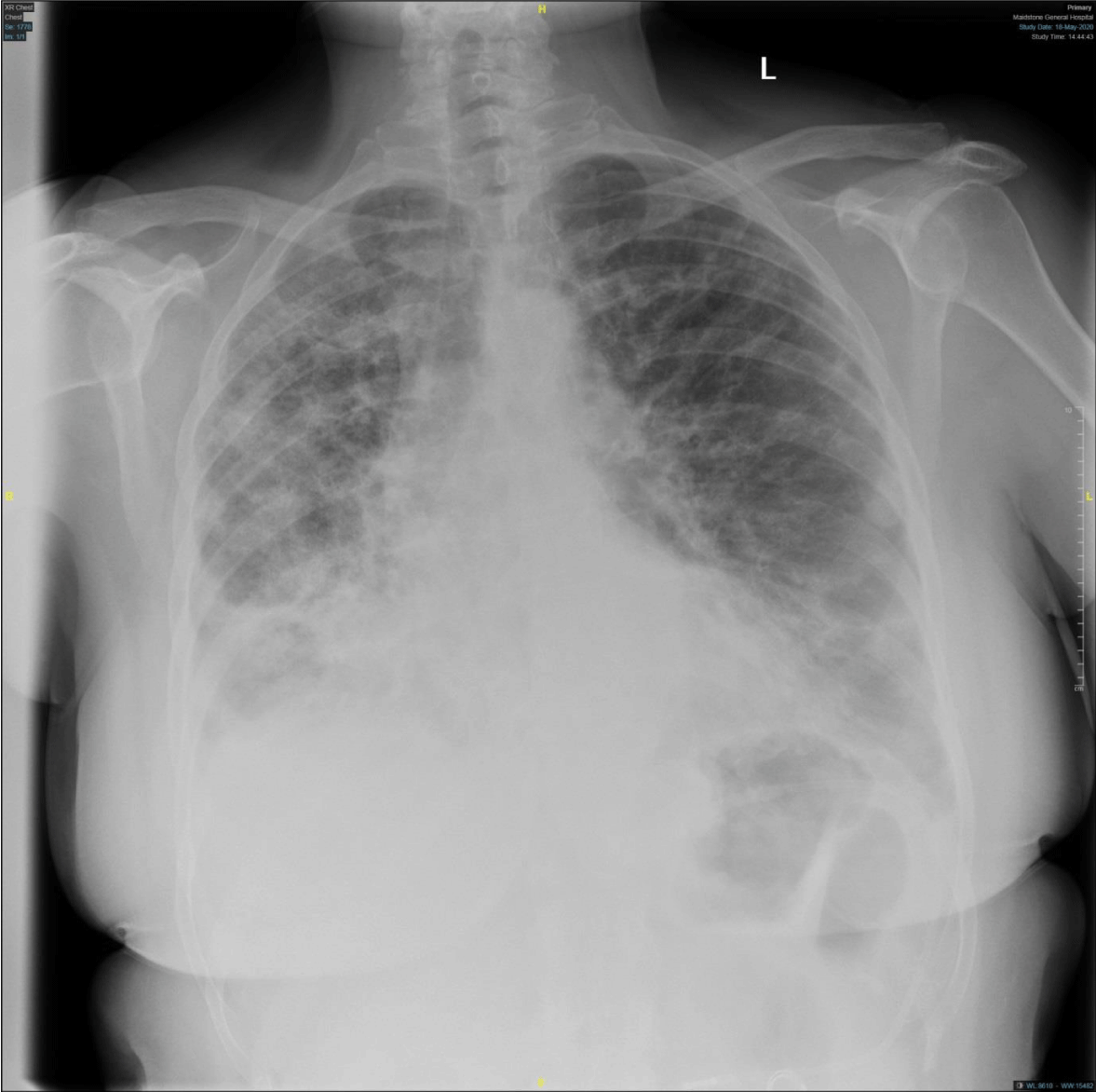
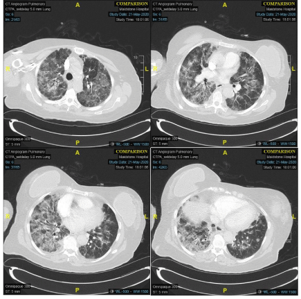
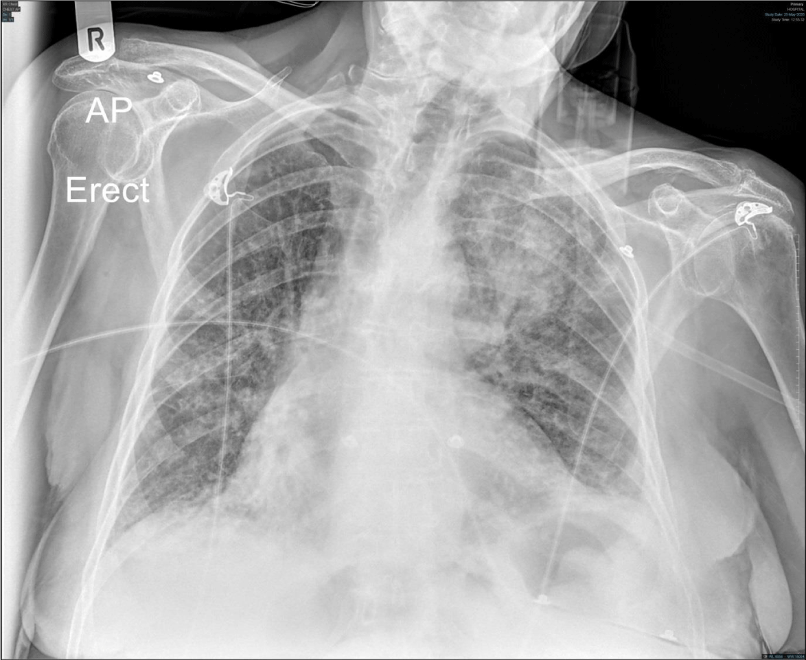
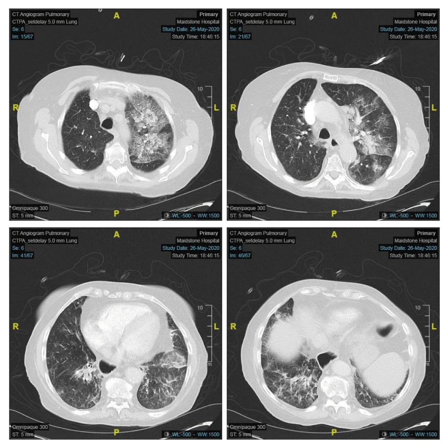
Given the concern raised by her clinical picture, inflammatory markers (Table 1) and a suspicious chest x-ray (Figure 1), she was initially treated as community acquired pneumonia but the high oxygen requirement and biochemical markers had prompted us to do a CT pulmonary angiogram (CTPA) which showed extensive bilateral ground glass changes felt to be consistent with COVID-19 (Figure 2). Due to persistent hypoxia despite standard oxygen therapy, she was commenced on CPAP.
During her admission, she had three serial negative RT-PCR COVID-19 swabs. However, the progression of her illness alongside high clinical suspicion led to a differential diagnosis of pneumonitis secondary to COVID-19 which ultimately prompted the use of steroids in addition to CPAP.
The patient ultimately improved after seven days of CPAP, a second course of antibiotics and a steroid wean. She was subsequently discharged home after making a full recovery.
Case 2
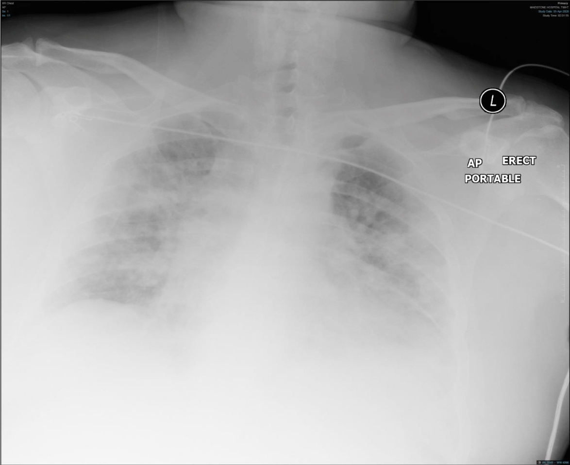
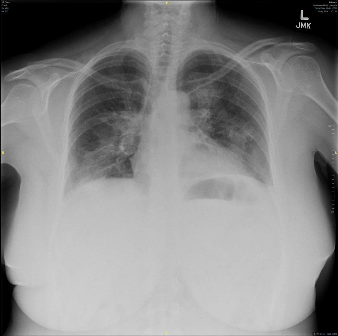
A 55-year-old male was admitted with pyrexia, cough and shortness of breath. He had a raised BMI (35) but was otherwise a fit, non-smoker. On admission, he was hypoxic and tachypnoeic; his chest x-ray (Figure 3) showed evidence of bilateral peripheral infiltrates and his inflammatory markers were elevated (Table 2).
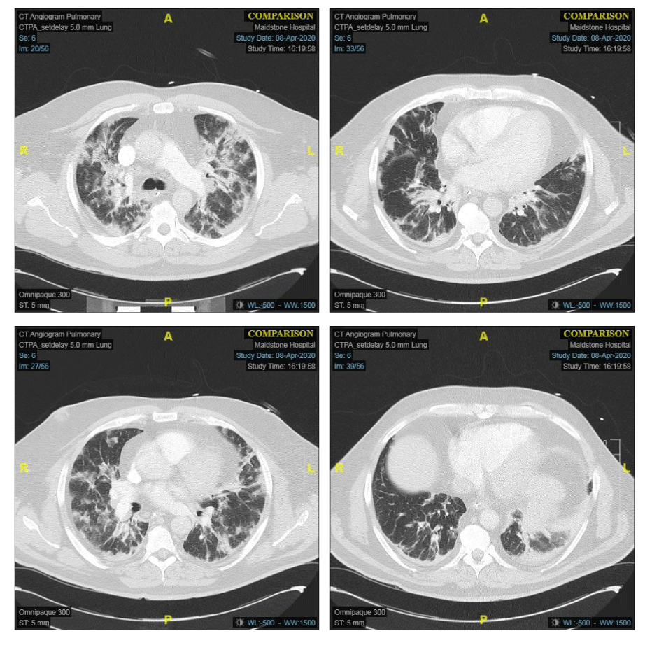
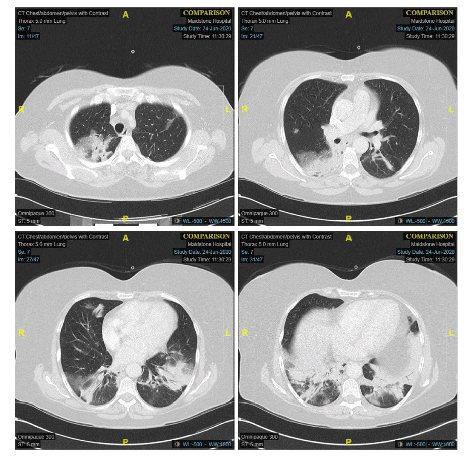
The patient had four negative serial PT-PCR COVID-19 swabs over a period of two weeks. He ultimately had a CTPA which showed bilateral peripheral consolidation with ground-glass opacification (Figure 4); this combined with his inflammatory response prompted a differential diagnosis of ARDS secondary to COVID-19.
The challenging issue was the location this patient was going to be treated due to his conflicting clinical picture.
He was managed actively with CPAP to maintain his oxygen requirement and his nutritional needs with an NG tube being placed with feeds. He required CPAP for 21 days & did not develop nosocomial infections during his stay. After extensive physiotherapy, he was discharged home with good recovery.
Case 3
A 79-year-old female presented with pyrexia, shortness of breath and a non-productive cough. She was known to suffer with frequent exacerbations of her COPD and bronchiectasis. Despite a history of heart failure, there was no evidence of decompensation preceding her admission. She was normally independent with a Rockwood clinical frailty score of 3.
Her chest radiograph showed extensive left upper lobe consolidation (Figure 5); this combined with her history and raised inflammatory markers (Table 3) led to an initial diagnosis of Community Acquired Pneumonia (CAP) on a background of emphysematous lung disease. She was treated with oral steroids, nebulisers and intravenous antibiotics. Alternative diagnoses such as atypical pneumonia or thromboembolic disease were considered to be a possible cause of her deterioration. During her admission, she had two RT-PCR COVID-19 swabs completed 48 hours apart both of which were negative.
The patient failed to improve and ultimately had a CTPA which showed extensive ground-glass opacity most confluent in the left upper lobe with focal consolidation (Figure 6). This imaging, alongside her biochemical and clinical picture, favoured a diagnosis of COVID-19 with secondary bacterial infection. The patient required a seven-day course of CPAP but ultimately made a good clinical recovery before discharge home.
Case 4
A 54-year female non-smoker presented with shortness of breath, pyrexia and a non-productive cough. On admission, she was hypoxic, tachycardic and pyrexial with raised inflammatory markers (Table 4). Her first RT-PCR swab was negative however her chest radiograph demonstrated bilateral infiltrates (Figure 7).
She developed type one respiratory failure which necessitated supplemental oxygen therapy with a maximum requirement of 40% FiO2. She also had a CTPA due to worsening hypoxia, which showed extensive areas of ground-glass changes (Figure 8). Given this in combination with her laboratory findings, she too was treated as a suspected COVID-19 case.
The patient was initially treated with antibiotics which were subsequently stopped due to a negative atypical screen, sputum and blood cultures. She was started on steroids for suspected pneumonitis from COVID-19 and ultimately made a rapid recovery with reduction in her oxygen requirements without the need for CPAP and was discharged home after making a good recovery.
Discussion
The cases described above show us that the universally accepted RT-PCR diagnosis is not entirely reliable on its own to exclude COVID-19 infection. If we were to rely on this alone, this would present a significant infection risk to both staff and other vulnerable patient groups due to the diagnostic dilemma. Existing reports suggest that there is only a 70% sensitivity of obtaining a definitive diagnosis from an RT-PCR swab versus a 98% diagnostic yield with CT scanning [1].
We believe this to in line with other authors due the diagnostic yield from the swab and its dependence on the following factors [2,3];
• Immature development of nucleic acid detection technology
• Variation in detection rate from different manufacturers
• Low patient viral load
• Improper clinical sampling: The reasons for the relatively lower RT-PCR detection rate in our sample compared to a prior report are unknown
• intrinsic limitations (i.e. collection and transportation of samples and diagnostic kit performance)
In view of the significant infectivity caused by COVID-19, a single or even multiple negative swabs does not necessarily dismiss the diagnosis especially in the presence of clinical, biochemical and radiological suspicion as demonstrated by the cases described above. Given the extensive and rapid spread of disease, more so when the R-value exceeds 1 [4], as well as the potential long term effects of the disease and associated therapies, we recommend that these aspects could supersede negative RT-PCR swabs in patients with high clinical suspicion.
As stated by other authors, if the pre-test probability is high, a CT scan would be useful to appropriately triage patients, ensure strict isolation protocols are maintained and treatment guided appropriately [5].
Several authors have already described that swab negative status does not rule out the possibility of COVID-19 infections; this has been described extensively and some have even recommended the use of bronchoalveolar lavage to aid in obtaining a diagnosis [6].
One of the factors that needs to be looked into and which would be extremely useful during the winter months which traditionally carry a high respiratory disease load would be to triage these patients appropriately and isolate away from other vulnerable groups of respiratory patients who have significantly impaired respiratory function and would do poorly in the event of a COVID-19 co-infection.
We also feel there is huge benefit in isolating these “Grey” cases away from the green zones dedicated for non-COVID infections, where people with advanced age, metabolic conditions and poor physiological reserve might succumb to the infection either due to the respiratory status or from the complications.
Learning Points
- Adopting Biochemical parameters at the front door for patients with respiratory symptoms: we recommend a COVID-19 blood test bundle including but not limited to: Full blood count, Urea, Electrolytes, Renal function, Liver function, Troponin, D-Dimer, Ferritin, Lactate Dehydrogenase, Pro-calcitonin, C-Reactive Protein
- CT is an important diagnostic aid in patients with diagnostic ambiguity and could supersede RT-PCR swabs [1,2,7].
Ideally, isolation in a negative pressure side room and the use of full respiratory precautions in these patients would be important not only to prevent them from infecting vulnerable cohorts but also to minimise the risk of them developing secondary infections themselves. This would also help protect healthcare workers and minimise any potential cross contamination between other patients.
- Fang Y, Zhang H, Xie J, Lin M, Ying L, Pang P, et al. Sensitivity of CT chest to R-PCR. Radiological society of North America. Radiology. 2020; 296: E115-E117. DOI: 10.1148/radiol.2020200432
- Yan Li, Liming Xia. Coronavirus Disease 2019 (COVID-19): Role of chest CT in diagnosis and management. American Journal of Roentgenology. 2020; 214: 1280-1286. DOI: 10.2214/AJR.20.22954
- Corona virus statistics and analysis, UK Gov Database, 2020. https://bit.ly/3iz0dXQ
- Song YG, Shin HS. COVID-19, A clinical syndrome manifesting as hypersensitivity pneumonitis. Infect Chemother. 2020; 52: 110-112. DOI: 10.3947/ic.2020.52.1.110
- Geoffrey D. Rubin , Christopher J. Ryerson, Linda B. Haramati, Nicola Sverzellati, Jeffrey P. Kanne, Suhail Raoof, et al. The Role of chest imaging in patient management during the COVID-19 pandemic: A multinational consensus statement from the Fleischner Society. Radiology. 2020; 296: 171-180. DOI: 10.1148/radiol.2020201365
- Winichakoon P, Chaiwarith R, Liwsrisakun C, Salee P, Goonna A, Limsukon A, et al Negative nasopharyngeal and oropharyngeal swabs do not rule out COVID-19. J Clin Microbiol. 2020; 58: 297-220. DOI: 10.1128/JCM.00297-20

Sign up for Article Alerts