Surgical Repair of Aorto-Ventricular Tunnel: A Case Report and Review of Methods?
Humberto Rodriguez1, Jesus Figueroa1, Jose Antonio Quibrera2, Luis Cordova3 and Thomas Di Sessa4*
1Hospital Pediatrico De Sinaloa Surgery Monterrey Mexico
2Hospital Pediatrico De Sinaloa Cardiology Culiacan Mexico
3Hospital Pediatrico De Sinaloa Antehesiology Culican Mexico
4Department of Pediatrics, University of Kentucky Lexington, Kentucky USA
*Address for Correspondence: Thomas Di Sessa, Department of Pediatrics, University of Kentucky Lexington, Kentucky USA, E-mail: [email protected]
Submitted: 17 January 2018; Approved: 18 January 2018; Published: 19 January 2018
Citation this article: Rodriguez H, Figueroa J, Quibrera JA, Cordova L, Di Sessa T. Surgical Repair of Aorto-Ventricular Tunnel: A Case Report and Review of Methods. Int J Cardiovasc Dis Diagn. 2018;3(1): 001-003.
Copyright: © 2018 Rodriguez H, et al. This is an open access article distributed under the Creative Commons Attribution License, which permits unrestricted use, distribution, and reproduction in any medium, provided the original work is properly cited
Keywords: Aorto-Left Ventricular Tunnel, Congenital heart Disease
Download Fulltext PDF
This is a case report outlining the anatomy of a rare congenital cardiac anomaly (aorto-left ventricular tunnel) and its surgical repair.
Introduction
Aorto-left ventricular tunnel is a rare congenital anomaly comprising only 0.1 to 0.5 % of congenital defects [1]. As of 2007 only 130 cases have been reported in the literature [1]. Anatomically it is an extracardiac connection between the right sinus of Valsalva either the left or right ventricle. Hovaguimian H et al has divided this anomaly into 4 types: type 1 – a slit like opening at the aortic end with no valve distortion 24 % of cases, type-2 associated with a large extracardiac aneurysm 44 % of cases, type - 3 associated with an intracardiac aneurysm of the septal portion of the tunnel with or without with or right ventricular outlet obstruction 24 % of cases, type - 4 a combination of 2 and 3 8 % of cases [2]. Physiologically it functions as aortic regurgitation producing left ventricular volume overload. Clinically most cases present in infancy with symptoms of refractory congestive heart failure. Occasionally patients present only with a heart murmur later in life. A variety of surgical approaches have been employed in the repair of the defect. We present herein a case of aorto-left ventricular tunnel successfully repaired at 7 days of age and a review the multiple surgical approaches.
Case Report
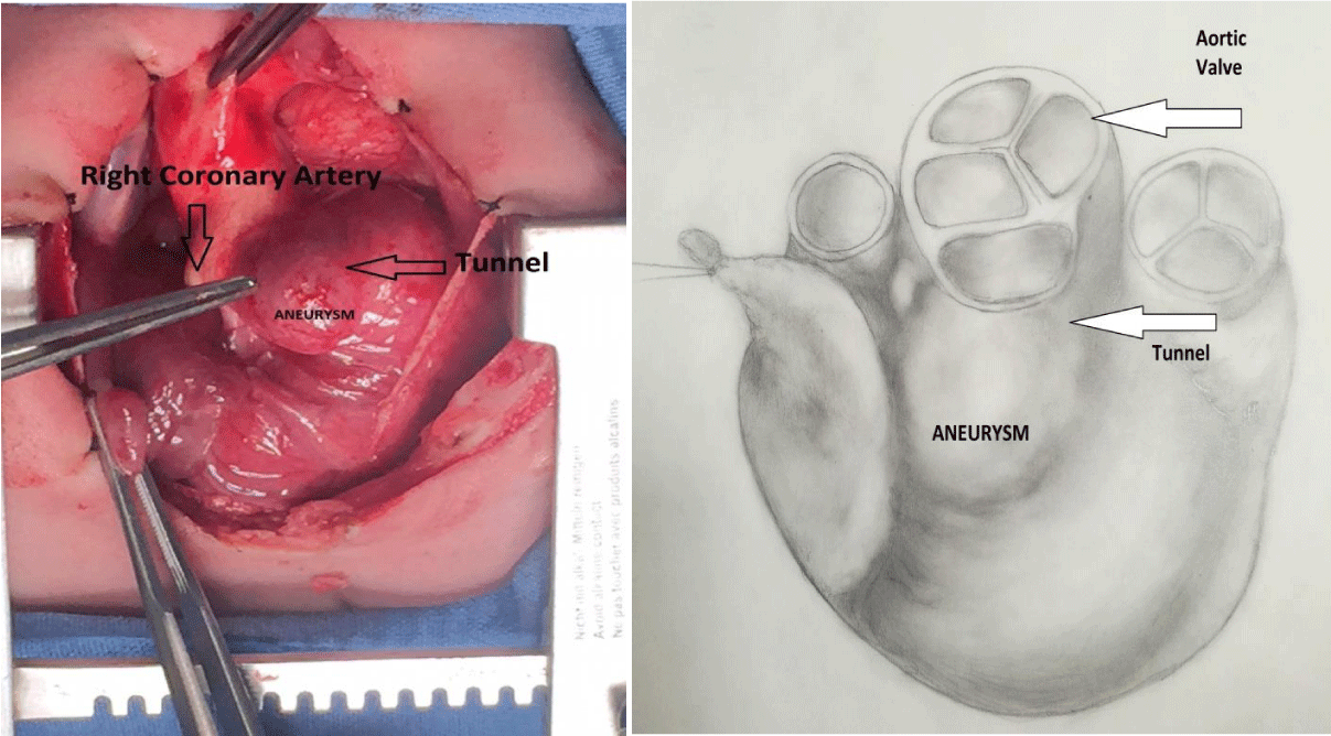
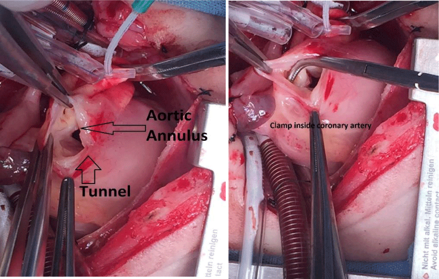
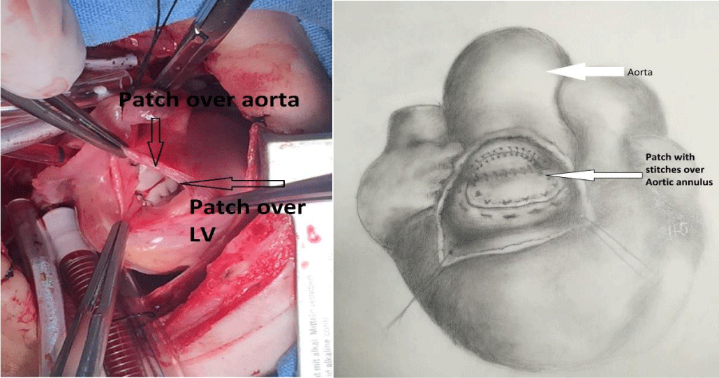
A one-day old male, product of a 39-week uncomplicated gestation, labor and delivery to 19-year-old female was noted to have respiratory distress characterized by intercostal retractions and nasal flaring shortly after birth. On physical examination there was a 3/6 long systolic murmur and a loud single second heart sound. Chest x-ray showed cardiomegaly. An echo-Doppler study demonstrated an atrial communication with left-to-right shunt, a ductus arteriosus with a bidirectional shunt, and a long communication between a dilated right sinus of Valsalva and left ventricle. There was antegrade flow into the aorta and retrograde into the left ventricle in diastole. Despite treatment with diuretics, the baby developed uncontrolled heart failure. The infant was taken to surgery on the seventh day of life. The procedure was performed with aortic cross-clamping, cardioplegia, ligature of both cavae and a right atriotomy. We observed a large bulbous aneurysm over the right sinus of Valsalva to the anterior Left Ventricular Outflow Tract (LVOT) (Figure 1 A and B). We used a longitudinal incision to open the aneurysm and found a communication between Aorta and LV, Aortic valve was normal and the right coronary artery was normal size and position (Figure 2A and B). The origin of the tunnel was above the sinotubular junction. The tunnel was closed along its anterior wall with a Goretex (need manufacturer) patch to close the aortic communication using a double running suture; the patch was extended to close LVOT orifice, using running suture over aortic annulus and around the LV orifice (Figure 3A and B). We closed remaining tissue of the aneurysm over the patch. The ASD and the atriotomy were repaired.
Discussion
This discussion will by no means provide the details of all types of surgical repair of aorto-left ventricular tunnel. Our goal is to briefly review the different approaches employed and their pros and cons. Experience has shown that for the best return of left ventricular size to normal repair should be before 6 months of age. There are a variety of techniques used to close an aorto-left ventricular tunnel (1). Adequate repair includes not only closure of the tunnel but also support of the aortic valve, maintaining normal coronary circulation as well as avoiding Left and right ventricular out obstruction. The following methods have been employed: 1) Closure of the aortic opening either by direct suture or with a patch; 2) closure of the ventricular end of the tunnel; 3) Obliteration of the Tunnel: and 4) closure of both orifices (3) In addition, a number of materials have been used as a patch (dacron, teflon, pericardium, piece of aneurysm) [3,4]. At the present time there appears to be debate regarding direct suture closure of the aortic end vs patch closure. Concern exists that direct suture closure of the aortic orifice distorts the aortic valve cusps and does not provide adequate support for the valve. Thus, patients may be more prone to long term development of aortic regurgitation [5]. However, Martins et al reported a 35-year experience with this anomaly. In this report 11 children had surgical repair. Six of nine patients repaired at their institution had direct aortic suture. No significant aortic regurgitation was observed at a mean follow up of 5 years [6]. Due to the rarity of this anomaly it is difficult to say which technique is preferred. Grunenfelder et al (4) state that patch closure of the aortic and ventricular openings is “the standard technique”. We chose this later approach using a single patch. This approach conformed to observed anatomy and complied with the objectives of repair. A not uncommon situation that occurs with aorto- left ventricular tunnel is origin of the right coronary artery along the length of the tunnel. Type of repair depends how distant along the tunnel the artery originates [1,4].
Conclusion
This case demonstrates the anatomy of the most common type of Aorto- left ventricular tunnel and a typical surgical approach. Moreover, we briefly discuss alternate surgical procedures.
- McKay R. Aorto-ventricular tunnel. Orphanet J of Rare Diseases. 2007; 2: 41. https://goo.gl/x6SQaB
- Hovaguimian H, Cobanoglu A, Starr. Aortico-left ventricular tunnel: a clinical review and new surgical classification. Ann Thorac Surg. 1988; 45: 106-112. https://goo.gl/jqyF8U
- Fotios A. Mitropoulos, Hillel Laks, Meletios A. Kanakis, Daniel Levi. Aorto-left ventricular tunnel: an alternative surgical approach. Ann of Thorac Surg. 2006; 82: 1113–1115. https://goo.gl/SeBQKQ
- Grunenfelder J, Zund G, Pretre R, Schmidi J, Vogt P, Turina M. Right coronary artery from aorto-left ventricular tunnel: case report of a new surgical approach. J Thorac Cardiovasc Surg. 1998; 116: 363-365. https://goo.gl/3muvZo
- Serino W, Andrade JL, Ross D, de Leval M Somerville J. Aorto-left ventricular communication after closure: late post-operative problems. Br Heart J. 1983; 49: 501-50. https://goo.gl/yUyMsC
- Martins JD, Sherwood, MC, Mayer JE, Keane JF. Aortico-left ventricular tunnel: 35-Year Experience. JACC. 2004; 44: 446-450. https://goo.gl/11DNSr


Sign up for Article Alerts