Which is More Important to Cerebral Blood Flow in Children with Vasovagal Syncope: Heart Rate or Blood Pressure??
Wang hong1*, Xing Yanlin1, Chen Rui1, Wang Ce1, Yu Xuexin1, Sun le1, Xu Yunming1, Li Xuemei1, Chu Yanqiu1 and Pan Xiaoli2
1Pediatric department of Shengjing hospital, China Medical University, Shenyang, China, 110004
2Neuro-function department of Shengjing hospital, China Medical University, Shenyang, China, 110004
*Address for Correspondence: Wang Hong, Associated professor, deputy director of Pediatric department of Shengjing hospital, China Medical University, Shenyang, China, Tel: 086-18940251677; E-mail: [email protected]
Submitted: 06 December 2017; Approved: 26 December 2017; Published: 28 December 2017
Citation this article: Hong W, Yanlin X, Rui C, Ce W, Xuexin Y, et al. Which is More Important to Cerebral Blood Flow in Children with Vasovagal Syncope: Heart Rate or Blood Pressure? Int J Cardiovasc Dis Diagn. 2017;2(3): 058-064.
Copyright: © 2017 Hong W, et al. This is an open access article distributed under the Creative Commons Attribution License, which permits unrestricted use, distribution, and reproduction in any medium, provided the original work is properly cited
Keywords: Cerebral Blood Flow; Syncope; Ambulatory Electroencephalogram; Heart Rate; Blood Pressure
Download Fulltext PDF
Objective: The mechanism of Vasovagal Syncope (VVs) is transient cerebral insufficient blood flow. It results in short unconsciousness and failed to maintain body posture. Once the cerebral blood flow decrease, the cerebral wave becomes slow. To exploring the effects of Heart Rate (HR) and Blood Pressure (BP) at syncope, we recorded the cerebral wave using Ambulatory Electroencephalogram (AEEG) in children during the Head-Up Tilt Test (HUTT).
Methods: Total 152 children from the pediatric department of Shengjing Hospital, China Medical University were enrolled between January 2008 and March 2016. Serum biomarker tests, Electroencephalogram (EEG), Transcranial Doppler (TCD) and head Magnetic Resonance Angiography (MRA) or head Computed Tomography (CT) was examined. HUTT was performed concomitantly with AEEG. The endpoint was undergoing the HUTT for 45 minutes or the occurrence of syncope/pre-syncope. Sublingual nitroglycerin provocative test (4-6 µg/kg, max 300 µg) (SNHUTT) was followed HUTT for 20 minutes or the occurrence of syncope/pre-syncope.
Results: AEEG was performed in 107 patients. Ninety one AEEG was synchronous with HUTT, while 16 AEEG performed before HUTT. Abnormal AEEG was found in 10 patients HUTT/SNHUTT positive in 9 patients. Among them four with symmetrical high amplitude slow wave were found in bilateral cerebral hemispheres at the moment of syncope/pre-syncope occurred during HUTT (3 vasodepressor and one mixed depressor) and disappeared as soon as they prostration. Four with unsymmetrical spike waves at sleeping and one with sleep disorder at no-syncope period. The other one was HUTT negative but with slow waves at all day. Eighty four patients were positive in HUTT/SNHUTT, while 18 were negative despite undergoing HUTT and SNHUTT. Vice Sinusitis was accidentally found on MRA in 10 patients. Four with vasodepressor, two mixed depressor, One Postural Tachycardia Syndrome (POTS), one undefined and two negative.
Conclusions: Hypotension may play a more important role than HR in cerebral hemodynamic when VVs occurred during HUTT. Sinusitis may induce VVs. AEEG is an important method to distinguish the syncope with unconsciousness or convulsion during HUTT.
Introduction
Syncope is a common disease. The most frequent cause of recurrent syncope is Vaso-Vagal Syncope (VVs) [1]. In order to give an exact diagnosis, the serum biomarkers, 12-lead EKG, AECG, PDE, EEG, AEEG, TCD, head MRA or CT need to performed to except hypoglycemia, cardiac syncope, central nerve systom disease. It was well know that HUTT is the gold standard to diagnose VVs [2]. Nearly 80 percent syncope children were diagnosed by HUTT. The mechanism of Vasovagal Syncope (VVs) is transient cerebral insufficient blood flow. It results in short unconsciousness and failed to maintain body posture. Once the cerebral blood flow decrease, the cerebral wave becomes slow. The aim of this study was to investigate effects of Heart Rate (HR) and Blood Pressure (BP) at syncope.
Materials and Methods
A total of 152 patients with syncope/pre syncope history were consecutively enrolled from the pediatric department of Shengjing Hospital, China Medical University, between January 2008 and March 2016. Exclusion criteria: cardiomyopathy, myocarditis, serious congenital heart disease, complex arrhythmia and cerebrovascular malformation. They underwent blood biomarkers test, included serum Hemoglobin (Hb), blood sugar, Creatine Kinase (CK), Creatine Kinase MB (CKMB), Creatine Kinase MB Mass (CKMB mass), Cardiac Troponin I (cTnI) and Highly Sensitive Cardiac Troponin T (hs-cTnT), ECG, AECG, PDE, EEG, TCD, head MRA or CT. HUTT or HUTT/SNHUTT were performed and synchronous recording AEEG.
HUTT Protocol and Endpoint
Each patient fasted for 8 hours, and 100 ml normal saline was prepared to inject intravenously in the left hand. AEEG was connected, a 12 lead ECG was placed, and the head-up tilt bed rising to 60-70 degrees. HR monitor for whole HUTT by Defibrillator. BP (right arm) and ECG lead II were recorded every minute for the first three minutes and then every five minutes for 45 minutes or syncope/pre-syncope occurred. If they agreement, the negative patients will continue to SNHUTT for another 20 minutes or syncope/pre–syncope occurred, while BP monitor and ECG lead II tracing every minute.
The criteria for syncope
Orthostatic Hypotension (OH): HR change less 10 bpm, SBP≤80 mmHg or DBP ≤ 50mmHg with syncope/pre-syncope performed within three minutes of HUTT. POTS: HR increase ≥ 40 bpm or over 130 bpm with syncope /pre-syncope within10 minutes of HUTT, at mean while SBP decrease less 20 mmHg and DBP decrease less 10 mmHg. Three subtype VVs were cardio depressor: HR < 75bpm in 4-6 years old, HR < 65bpm in 7-8 years old, HR < 60bpm over 8 years old; SBP without significant change; Vasodepressor: HR without significant changed, SBP≤80 mmHg or DBP ≤ 50mmHg with syncope or pre-syncope; Mixed depressor: both HR and BP decrease.
Instruments and Equipments
ECG: NIHON KOHDEN 91309, Japan. ECG monitoring: LIFEPAK 20 Defibrillator, Physio-Control, US Monitor: PM-7000, Shenzhen Mairui Biological Medical Electronic co., LTD, China. AECG: TLC3000B, Qinhuangdao Conde Medical Systems co., LTD, China. TCD: DWL, Germany. EEG: Medelec, England. AEEG: 32 leads Voyageur, Nicolet, US. CT: Toshiba 64 spiral CT, Japan. MRA: imaging of nuclear magnetic resonance (NMR) and artery angiography, Intera Achieva 3.0T, PHILIPS Netherlands. Bed of HUTT: Beijing Juchi Pharmaceutical Co., LTD, China.
Statistics analysis
Categorical data was shown with means ± standard deviation. The comparison of means was performed with t test. The rate was compared using the χ2 test. Data analysis was conducted with SSPS11.5 software and P < 0.05 was considered to be a significant difference.
Results
General information in children with syncope
Table 1 was the summary of general information in children with syncope. Hb in female is lower than male, P<0.05.
Clinic examination results in syncope children
Table 2 showed the examination results in syncope children. In one boy with left ventricle hypertrophy on ECG but normal anatomy and systolic function of the left ventricle on PDE, BP and AECG before HUTT, the heart rhythm changed from sinus to atrio-ventricular node when pre-syncope occurred during HUTT. His lowest HR was 39 bpm and resolved once he lay on his back. Two children with sinus bradycardia on AECG, one with mixed depressor and the other one with uncertain in HUTT. A male with paroxysmal sinus tachycardia on AECG had vasodepressor in HUTT.
Total of nine patients HUTT positive with abnormal AEEG: 4 with slow waves on AEEG at the moment of syncope or pre-syncope occurred during HUTT, which 3 showed 2-3 Hz high-voltage slow waves and one showed 5-7 Hz, 4 with spikes and discharge wave on AEEG at sleeping and one had sleep disturbance waves. But all 5 were without a corresponding clinic events during HUTT. One case HUTT was negative but with 2-3 Hz slow wave on EEG and AEEG. His MR suggests encephalitis with the bilateral temporal parietal lobe increased T2W1 signal.
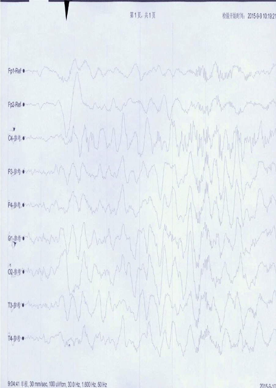
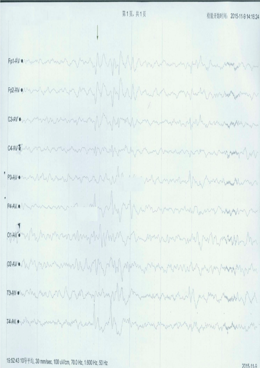
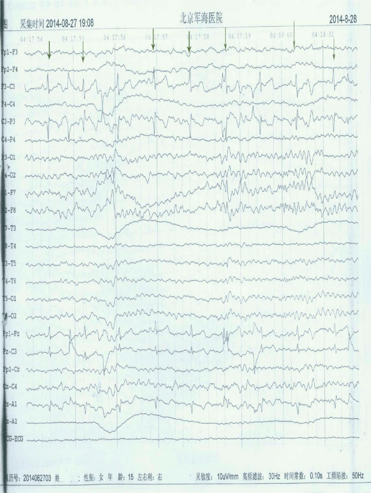
In TCD, 11 patients had rapid cerebral blood flow but normal EEG and AEEG. All 16 patients head CT were normal. Of 111 children underwent head MRA, 25 had abnormal results: 10 with paranasal sinusitis (4 vaso-depressor, 2 mixed-depressor, one POTS, one uncertain and 2 negative on HUTT). 5 cerebral vassal stenosis (3 vertebral artery, 2 arteriae cerebri anterior); 5 with arachnoid cyst of left temporal pole; 3 with outside the brain rests slightly widened, one with cyst of pellucid septal cave; one with increased T2W1 signal in bilateral parietal temporal lobe. A vaso depressor girl with transient face and limb convulsions, her AEEG showed transient high amplitude discharges of bilateral cerebral hemispheres with high 2-3 Hz slow waves (Figure 1); A boy with frequent syncope but normal AEEG during faint when HUTT, while epileptic waves was recorded during sleep on AEEG. This wave has no diagnostic value but would be useful on follow-up (Figure 2). Another girl had multiple episodes of syncope without twitch occurred. Two weeks ago she took video EEG in local hospital and found epileptic waves during sleeping (Figure 3). Her parents didn’t agree to undergo AEEG during HUTT. Followed HR and BP goes down, she had a faint and transient facial convulsion lasting 3-4 seconds and resolved after laying down on her back without accompanying urinary incontinence. MRA usually be use to find congenital malformation of cerebral artery. A POTS girl, with Kawasaki disease five years ago, was found left vertebral artery stenosis (near closure) in MRA but normal AEEG during syncope occurred, it suggests that her syncope was no relation to her structural change.
HUTT results
Table 3 was the summary of HUTT results. Forty seven of 152 HUTT positive and 37/55 HUTT negative children were positive after SNHUTT. The males were elder than females, showed significant difference, P < 0.05.
A patient in this study was hospitalized two times for syncope recurrent, initial testing showed POTS and half year later his HUTT showed VVs vasodepressor type. After talking with his father we learn that his parents had divorced, he lived with his father and a stepbrother. He possibly felt that he was treated coldly. He frequent fainted after his father remarried. After he consulted a psychologist his syncope was never happened again.
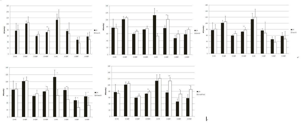
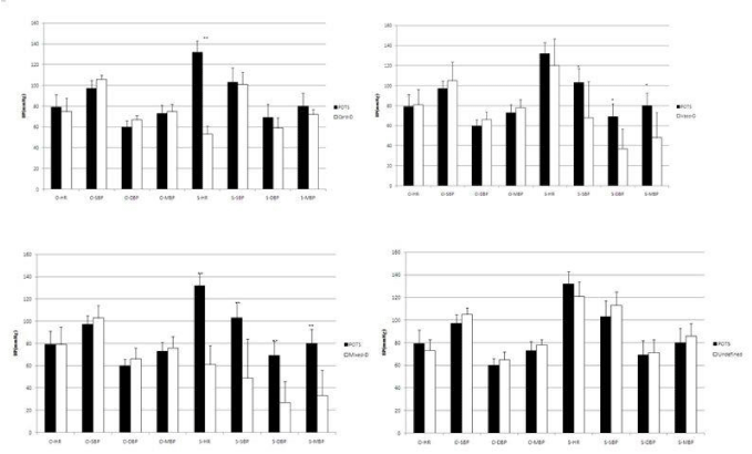
Table 4 and figure 4-7 showed the comparison between different types of syncope with HUTT.
A boy with pseudo-syncope, his syncope occurred with normal HR and BP during HUTT.
Discussion
It has been reported that over half syncope patients of teenage were benign [3]. According to an investigation in Beijing and Hunan and Hubei provinces, VVs was the most common type among children [4]. This study showed similar results. At the beginning of HUTT, 13 children with syncope or pre-syncope after three minutes had SBP increased over 20 mmHg and/or MAP elevated over 10 mmHg. It was unclear they should belong to which types and we named them as ‘undefined ’. Someone with syncope/pre-syncope with HR over 130 bpm or HR increased over 40 bpm that occurred 10 minutes later after HUTT and couldn’t be diagnosed POTS. We found that if HUTT is continued, their BP or both BP and HR dropped and should belong to VVs. Further talking about with the pseudo-syncope boy’s father we learned he has an overcritical mother. After consultation with a psychologist he was diagnosed with body conversion disorder and hysteria [5]. He improved after six months of psychological counseling and treatment with Sertraline. Typically psychogenic pseudo-syncope presents in a pediatric patient with excessive parental monitoring as well as negative self-image [6]. Among 97 patients over three years in a pediatric cardiology syncope center in France, 70.4% were VVs, 20.6% were psychogenic pseudo-syncope and 6.2% were cardiogenic syncope [7]. Of course, if syncope occurs with action or anger, potentially life-threatening disease could be the cause [8,9].
It was well known that deceasing cerebral blood flow can result syncope [10]. Transcranial Doppler (TCD) was the best way to monitor cerebral blood flow [11], but it is inconvenience to operate during HUTT when the bed rising and down [12]. Cerebritis children maybe accompany syncope and they can be found slow wave at any time on EEG, while VVs only transient slow wave and epilepsy outbreak is spike and ware wave on EEG. Therefore, with AEEG to monitor brain waves can distinguish them very well when syncope occurred during HUTT. The AEEG change of image one was in agreement with the report that there was insufficient blood supply in upright syncope [13]. For the episode wasn’t monitored with AEEG in patient image 3, epilepsy couldn’t be except. In this study, cardiac depressor/ mixed depressor’s HR decreased to criteria and recovered as soon as they are prostrate, while some vasodepressor/mixed depressor kept their SBP/DBP/MBP lower or continue goes down for 1 to 2 minutes after prostration, then gradually recovered, such as these 4 patients with slow waves during syncope. Therefore, we deduced the blood pressure may play a more important role than HR in VVs. Thus we should stop HUTT as soon as the syncope or pre syncope occurred and the BP decreasing reached diagnostic criteria to prevent brain damage, furthermore, preparing venous access and normal saline before HUTT was essential.
The syncope type could be variable. VVs related to emotion and children with frequent syncope usually have some psychosocial stressors [14]. Psychological consulting in addition to medication may be useful. According to a recent report, syncope type can be predicted from HR and BP at the beginning of HUTT [15]. That is good news to those unwilling to undergo provocative testing. Among those various treatments for VVs, once felt pre-syncope, changing position to alleviate symptoms of dizziness [16] is a most convenience method. In this study, the basic HR of males was faster than females in those who underwent SNHUTT. This may be due to the fact that boys have a greater degree than girls of sympathetic nervous system excitement before SNHUTT [17]. A recent study suggested that antihypertensive medication might be better than a pacemaker in hypertensive patients with vasovagal hypotension [18]. For many children with VVs have a positive family history, genetic screening for Gaussian protein maybe helpful to diagnose vasovagal syncope.
In this study, total 9 with vice sinusitis was on head MR and 7 HUTT were positive, we couldn’t find related paper to explain it, maybe vice sinusitis stimulate cerebral artery and result them transient contract?
In summary, VVs is a common disease in teenage, the syncope type is variable. As soon as the patient syncope/pre syncope happened and his/her HR and/or BP (especially BP) meet the criteria of VVs, the HUTT should be stop and prepare normal saline to intravenous infusions. Vice sinusitis may reduce VVs.
Acknowledgement
This paper is funded by the Science and Technology Agency of Liaoning province (No. 2013225089)
- Vaddadi G, Corcoran SJ, Esler M. Management strategies for recurrent vasovagal syncope. Intern Med J. 2010; 40: 554-60. https://goo.gl/Q3yeiT
- Smith JA, Karalis DG, Rosso AL, Grothusen JR, Hessen SE, Schwartzman RJ, et al. Syncope in complex regional pain syndrome. Clin Cardiol. 2011; 34: 222-225. https://goo.gl/yPoj9m
- Naranje KM, Bansal A, Singhi SC. Fainting Attacks in Children. Indian J Pediatr. 2012; 79: 362-6. https://goo.gl/qi3TWx
- Chen L, Wang C, Wang H, Tian H, Tang C, Jin H, et al. Underlying diseases in syncope of children in China. Med Sci Monit. 2011; 17: 49-53. https://goo.gl/x2sgsA
- Bat T, Collins KK, Schaffer MS. Syncope during exercise: just another benign vasovagal event? Curr Opin Pediatr. 2011; 23: 573-5. https://goo.gl/KdgzkK
- Romme JJ, van Dijk N, Go-Schon IK, Casteelen G, Wieling W, Reitsma JB, et al. Association between psychological complaints and recurrence of vasovagal syncope. Clin Auton Res. 2011; 21: 373-80. https://goo.gl/Gkwia5
- Rio-Casanova LD, González A, Paramo M. Excitatory and inhibitory conversive experiences: neurobiological features involving positive and negative conversion symptoms. Rev Neurosci. 2016; 27: 101-110. https://goo.gl/8R2FvP
- Romme JJ, van Dijk N, Go-Schon IK, et al. Effectiveness of midodrine treatment in patients with recurrent vasovagal syncope not responding to non-pharmacological treatment (Stand-trial). Europace. 2011; 13: 1639-47. https://goo.gl/wvtXwc
- Cheng R, Shang Y, Wang S, Evans JM, Rayapati A, Randall DC, et al. Near-infrared diffuse optical monitoring of cerebral blood flow and oxygenation for the prediction of vasovagal syncope. J Biomed Opt. 2014; 19: 17001. https://goo.gl/US9p9f
- Norcliffe-Kaufmann L, Galindo-Mendez B, Garcia-Guarniz AL, Villarreal-Vitorica E, Novak V. Transcranial Doppler in autonomic testing: standards and clinical applications. Clin Auton Res. 2017; 18. https://goo.gl/kmi4ez
- Alboni P, Alboni M. Typical vasovagal syncope as a "defense mechanism" for the heart by contrasting sympathetic overactivity. Clin Auton Res. 2017; 27: 253-61. https://goo.gl/xvEQo9
- Blanc JJ, Benditt DG. Vasovagal Syncope: Hypothesis Focusing on Its Being a Clinical Feature Unique to Humans. J Cardiovasc Electrophysiol. 2016; 27: 623-9. https://goo.gl/Wt49CQ
- Del Rio-Casanova L, Gonzalez A, Paramo M, et al. Emotion regulation strategies in trauma-related disorders: pathways linking neurobiology and clinical manifestations. Rev Neurosci. 2016; 27: 385-95. https://goo.gl/jPGD57
- Ho D, Ghods M, Kumar S, Warrier N, Ilias Basha H, Budzikowski AS, et al. Early Hemodynamic Changes during Head-Up Tilt Table Testing Can Predict a Neurocardiogenic Response in an African-American Patient Population. Cardiology. 2016; 133: 223-32. https://goo.gl/Hb8yXn
- Reulecke S, Charleston-Villalobos S, Voss A, et al. Temporal analysis of cardiac autonomic regulation during orthostatic challenge by short-term symbolic dynamics. Conf Proc IEEE Eng Med Biol Soc. 2015; 2015: 2067-70. https://goo.gl/g3ZbB3
- Reulecke S, Charleston-Villalobos S, Voss A, Gonzalez-Camarena R, Gonzalez-Hermosillo J, Gaitan-Gonzalez MJ, et al. Men and women should be separately investigated in studies of orthostatic challenge due to different gender-related dynamics of autonomic response. Physiol Meas. 2016; 37: 314-32. https://goo.gl/cp1rD9
- Sutton R. Syncope in Patients with Pacemakers. Arrhythm Electrophysiol Rev. 2015; 4: 189-92. https://goo.gl/sTZKhr
- Lelonek M, Zelazowska M, Pietrucha T. Genetic Variation in Gsα Protein as a New Indicator in Screening Test for Vasovagal Syncope. Circ J. 2011; 75: 2182-6. https://goo.gl/jDQMjW


Sign up for Article Alerts