Acute Coronary Syndrome Resulting From Compression of Left Main Coronary Artery Secondary to Pulmonary Aneurysm Expansion?
Jiang Y. Wang, Han Chen*, Xi Su, Chen W. Liu, Dan Song, and Hua Yan
Department of cardiology, Wuhan Asia heart hospital, Wuhan, China
*Address for Correspondence: Han Chen, Department of cardiac surgery, Wuhan Asia heart hospital, Wuhan 430022, China, Tel: +8615172496706; E-mail: 252810036 @qq.com
Submitted: 23 September 2017; Approved: 05 October 2017; Published: 07 October 2017
Citation this article: Wang JY, Chen H, Su X, Liu CW, Song D, et al. Acute Coronary Syndrome Resulting From Compression of Left Main Coronary Artery Secondary to Pulmonary Aneurysm Expansion. Int J Cardiovasc Dis Diagn. 2017;2(3): 055-057.
Copyright: © 2017 Chen H, et al. This is an open access article distributed under the Creative Commons Attribution License, which permits unrestricted use, distribution, and reproduction in any medium, provided the original work is properly cited
Download Fulltext PDF
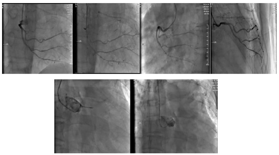
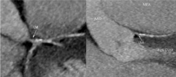
A 51-year-old man presented to the emergency department with intermittent, nonexertional chest pain of 10 days’ duration. Symptoms of chest pain were aggravated when the patient was on the left side. At 18 years of age, he had been diagnosed with a “hole in his heart” in the local hospital. His parents had declined options for treatment. On the Electrocardiogram (ECG) at emergency room, signs of acute myocardial infarction in the anterior territory were presented with ST depression in leads V1 to V6 and elevated in leads AVR, I, II. Troponin I concentration was 3.28 ng per milliliter (reference range, 0 to 0.03ng per milliliter). After stabilization in the emergency department, he was transferred to coronary care, where echocardiography demonstrated a persistent Atrial Septal Defect (ASD) measuring 4.3×3.6 cm with a right-to-left shunt consistent with severe pulmonary hypertension and Eisenmenger syndrome. Left ventricular function was severely impaired (ejection fraction=35%). Following a review by the cardiology team, 5-F multipurpose catheter was repeatedly tried into the left and right coronary sinus successful, angiography showed 90% stenosis in proximal left main coronary artery stent and 50% lesion in the proximal left circumflex artery, and known occluded left anterior descending coronary artery with right to left collaterals (Figure 1). Multi detector computed tomography showed central pulmonary artery aneurysms; the main pulmonary trunk was 71.0 mm, the left pulmonary artery 45.3 mm, and the right pulmonary artery 66.2mm; the left pulmonary artery, and the basal segment of the pulmonary artery wall thrombus, accompanied by the calcification; the left pulmonary arterial trunk thrombosis was about 12.4mm (Figure 2). Coronary artery CT image showed the left main coronary artery was compressed arc depression, and the lumen stenosis is greater than 70% (Figure 3). Ultimately, the patient was executed to LPA thrombology +LPA endarterectomy + pulmonary artery angioplasty +ASD repair +TVP+FO seam closure + left main coronary artery exploration + temporary pacemaker resettlement. He was discharged to home 14 days after her presentation. Post-cardio surgical operation coronary artery CT image showed left main coronary artery was not narrowed (Figure 4).
Compression of the left main coronary trunk has not been widely known. However, in 1957, Corday et al. [1], first pointed out that the distended main pulmonary artery easily compressed the left main coronary trunk because of the spatial position of the proximal part of the left coronary artery closed to the main pulmonary artery. Schaffer et al. [2], tried to prove the correlation between pulmonary pressure and coronary flow at autopsy which, however, was unsuccessful because the main pulmonary artery of the autopsied heart was not sufficiently dilated. Mitsudo et al. [3], first reported the case of a female patient with ASD that was combined with severe narrowing of the ostium of the left main coronary trunk on angiography. After the patient died suddenly, the postmortem examination confirmed no anatomic or pathological abnormality of the coronary arteries. Also, they reported that, in adults with ASD and pulmonary hypertension, the coronary angiography revealed localized concave narrowing of the left main coronary trunk orifice in 7 (44%) of 16 patients. In patients without pulmonary hypertension, however, these findings were not observed. Fijiwara et al. [4], reported 3 cases of adult ASD with compression of the left main coronary trunk by dilated main pulmonary artery. In one patient of the three, magnetic resonance imaging study demonstrated that the left main coronary trunk ran close to the left main pulmonary artery so that the markedly distended pulmonary artery easily compressed the left main coronary trunk. In two of the three cases, one case died because of septicemia after closure of ASD, the narrowed left main coronary artery was no longer narrowed or improved the severity of narrowing after closure of ASD. In our case, pulmonary hypertension due to ASD had already been present before the corrective surgery and it was completely improved after the correction of the shunt up to the current admission. We concluded that the dilated pulmonary artery trunk due to ASD associated with pulmonary hypertension can be a possible cause of left main coronary artery occlusion and that, if the ASD closure was executed, the narrowing or occlusion of the left coronary artery could be resolved, even after operation, owing to irreversible pulmonary hypertension.
- Corday E, Gold H, Kaplan L. Coronary artery compression. Trans Am Coll Cardiol. 1957; 7: 93–103. https://goo.gl/EqXCa7
- Schaffer Al, Bonaccorci B, Tchertkoff C. Compressibility of the coronary artery by the pulmonary artery distension. Am J Cardiol. 1963; 12: 406–7. https://goo.gl/JKHYgk
- Mitsudo K, Fujino T, Matsunaga K, Doi O, Nishihara Y, Awa J, et al. The coronary angiographic findings in the patients with atrial septal defect and pulmonary hypertension (ASD+PH): Compression of the left main coronary artery by pulmonary trunk (in Japanese). Kokyo to Junkan. 1989; 37: 649–655. https://goo.gl/6LGbwg
- Fujiwara K, Naito Y, Higashiue S, Takagaki Y, Goto Y, Okamoto M, et al. Left main coronary trunk compression by dilated main pulmonary artery in atrial septal defect. J Thorac Cardiovasc Surg. 1992; 104: 449–452. https://goo.gl/zrXAWa


Sign up for Article Alerts