Spontaneous Abdominal Hematoma in a Patient Treated With Ticagrelor and Aspirin?
Jiang Y. Wang, Xi Su*, Chen W. Liu, Chen YiXu, Zhi P. Zhang, Dan Song, JianPeng and Hua Yan
Department of Cardiology, Wuhan Asia heart hospital, Wuhan, China
*Address for Correspondence: Xi Su. Department of Cardiology, Wuhan Asia heart hospital, Wuhan 430022, China, Tel: +8615172496706; E-mail: [email protected]
Submitted: 22 September 2017; Approved: 29 September 2017; Published: 30 September 2017
Citation this article: Wang JY, Su X, Liu CW, YiXu C, Zhang ZP, et al. Spontaneous Abdominal Hematoma in a Patient Treated With Ticagrelor and Aspirin. Int J Cardiovasc Dis Diagn. 2017;2(3): 052-054.
Copyright: © 2017 Wang JY, et al. This is an open access article distributed under the Creative Commons Attribution License, which permits unrestricted use, distribution, and reproduction in any medium, provided the original work is properly cited
Download Fulltext PDF
Spontaneous Retroperitoneal Hematoma (SRH) is a potentially fatal and difficult to diagnose clinical entity defined as bleeding into the retroperitoneal space without associated trauma or iatrogenic manipulation. We present a case of spontaneous retroperitoneal hemorrhage after acute myocardial infarction involving anticoagulant agents.
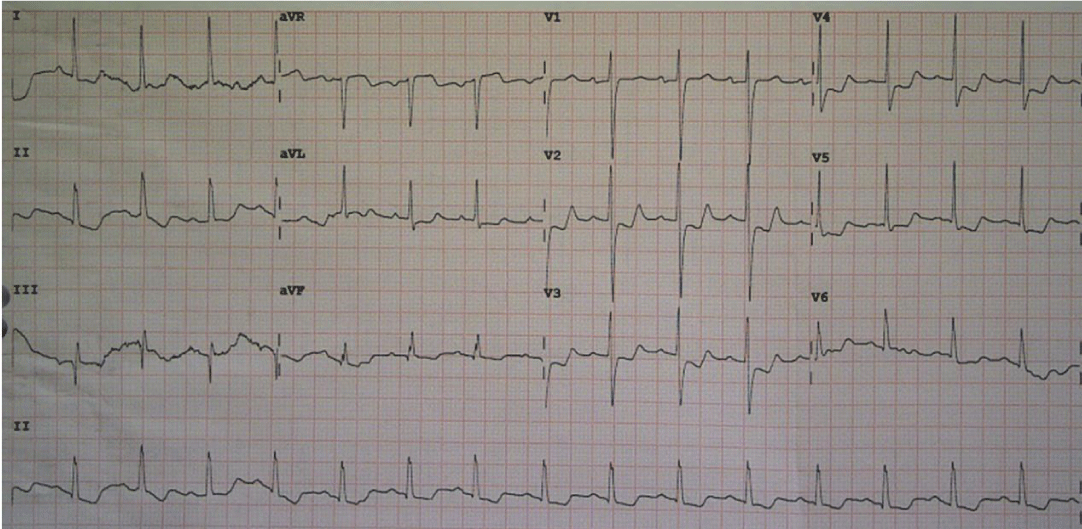
We introduce a case of a 71-year-old woman with a clinical history of hypertension, who presented to the emergency department with chest pain of 3-day duration. On the Electrocardiogram (ECG) at emergency room, signs of left main (LM) or Proximal Left Anterior Descending (LAD) stenosis were presented with ST depression in leads I,II,AVL,AVF,V1 to V6 and ST elevation in lead AVR (Figure.1). Troponin I concentration was 0.324 ng per milliliter (reference range, 0 to 0.04 ng per milliliter) and hemoglobin concentration of 126g/L. The transthoracic echocardiogram demonstrated no ventricular wall motion abnormalities, aortic valve mild regurgitation, no signs of aortic dissection. Emergency personnel considered the diagnosis of acute myocardial infarction with routine protocol for NSTEMI including loading doses of aspirin (300mg), ticagrelor (180mg) and rosuvastatin (20mg) followed by intravenous injection of unfractionated heparin (5000U). Then he was transferred to the coronary care unit. Following a review by the cardiology team, the emergency coronary angiogram (the right radial artery path) revealed isolated critical stenosis of the ostial-LM and mid-LAD and total occlusion of RCA (Figure.2A, C). He is performed emergency percutaneous coronary intervention. The LM and LAD stenosis was successfully treated with a drug-eluting stent, respectively (Figure.2B).
Then he was transferred to the coronary care unit. While the patient experienced extreme abdominal pain, nausea and vomiting (gastric contents, no bloody substance) at the time of coronary care unit. At the time his heart rate 115 beats/min, blood pressure 78/43mmHg, there was not a thoracoabdominal bruit and hypoactive bowel sounds. The transthoracic echocardiogram demonstrated no cardiac mechanical complications. Abdominal ultrasound demonstrated no retroperitoneal hematoma. Electrocardiogram demonstrated no evidence suggests that myocardial ischemia. Troponin I concentration was 0.892 ng per milliliter and hemoglobin concentration of 79g/L. He initially was diagnosed as hemorrhagic shock, and we decided to perform blood transfusions. Based on current clinical evidence, the evidence was not sufficient diagnosis of gastrointestinal bleeding. Immediately following, on the abdominal computed tomography, signs of retroperitoneal hematoma were presented with bilateral perirenal fascia and peritoneum slightly thickened, bilateral iliac muscle swelling (Figure.3A-E). After rehydration, blood transfusion, reversal of anticoagulation therapy and hemodynamic stability, and the patient was discharged home on day 7.
Falling, crush injury, road traffic accidents, injury to the retroperitoneal organs, fracture of the pelvis or lower spine, and retroperitoneal vascular injury are all potential causes of retroperitoneal hemorrhage or hematoma. Spontaneous retroperitoneal hematoma is a much less common distinct clinical entity that can be life-threatening [1]. Previous accounts consist mostly of small case-based series or single case reports that usually highlight the relationship between SRH and the use of antiplatelet or anticoagulant therapies [2-7].However, SRH is a more complicated disease, with considerable heterogeneity. SRH is an uncommon clinical entity that can lead to serious complications if not recognized and treated immediately. Patients with SRH often presented in a hypovolemic state with pain in the abdomen, back, legs, or hips [8]. There was a strong but not absolute relationship between SRH and previous treatment with blood-thinning medications [8]. Management consisted of aggressive resuscitation and reversal of coagulopathy or interventional radiology procedures [8]. Physicians are encouraged to suspect SRH in patients with centrally located pain and signs of hypovolemia, regardless of anticoagulation or antiplatelet history.
In conclusion, SRH is an uncommon but potentially lethal entity with a non-specific presentation that can lead to misdiagnosis and delayed treatment. It could be suspected in patients who present with truncal pain or hypovolemia-related symptoms, regardless of anticoagulation or antiplatelet history. CT imaging was frequently used and effective for diagnosis. The majority of patients received intensive-care-unitlevel of care and aggressive support in the form of blood transfusion, reversal of coagulopathy, or procedures by interventional radiology. Surgery was rarely indicated.
Acknowledgements
This study was supported by a grant from Wuhan Health and Family Planning Commission Research Foundation funded (Grant No.WX17Q36).rarely indicated.
- Rapp N, Audibert G, Gerbaud PF, Grosdidier G, Laxenaire MC. Spontaneous retroperitoneal hematoma: a rare cause of hemorrhagic shock. Ann Fr Anesth Reanim 1994; 13: 853-6. https://goo.gl/UXHfC2
- González C, Penado S, Llata L, Valero C, Riancho JA. The clinical spectrum of retroperitoneal hematoma in anticoagulated patients. Medicine (Baltimore) 2003; 82: 257-62. https://goo.gl/dYPX3v
- Jiang J, Ding X, Zhang G, Su Q, Wang Z, Hu S, et al. Spontaneous retroperitoneal hematoma associated with iliac vein rupture. J Vasc Surg 2010; 52: 1278-82. https://goo.gl/CPXvob
- Lissoway J, Booth A. Fatal retroperitoneal hematoma after enoxaparin administration in a patient with paroxysmal atrial flutter. Am J Health Syst Pharm 2010; 67: 806-9. https://goo.gl/Chg8qa
- Otrock ZK, Sawaya JI, Zebian RC, Taher AT. Spontaneous abdominal hematoma in a patient treated with clopidogrel and aspirin. Ann Hematol 2006; 85: 743-4. https://goo.gl/AhstFu
- Carrilero Zaragoza G, Egea Valenzuela J, Moya Arnao M, Muñoz Tornero M, Jijon Crespin R, Tomas Pujante P, et al. Spontaneous retroperitoneal hematoma in a patient under anticoagulant agents presenting as upper gastrointestinal bleeding. Rev Esp Enferm Dig 2016; 108: 817-818. https://goo.gl/6eBLBb
- Simsek A, Ozgor F, Yuksel B, Bastu E, Akbulut MF, Kucuktopcu O, et al. Spontaneous retroperitoneal hematoma associated with anticoagulation therapy and antiplatet therapy: two centers experiences. Arch Ital Urol Androl 2014; 86: 266-9. https://goo.gl/E2MGxD
- Sunga KL, Bellolio MF, Gilmore RM, et al. Spontaneous retroperitoneal hematoma: etiology, characteristics, management, and outcome. J Emerg Med 2012; 43: e157-61. https://goo.gl/XPexyY


Sign up for Article Alerts