Fat in the Coronary Arteries Isn’t Always a Villain?
Carlos A. Espinoza*, Syed Ali, Lavanya Alapati and Ashesh N. Buch
Interventional Cardiology Fellow, Department of Cardiovascular Sciences, East Carolina Heart Institute at East Carolina University
*Address for Correspondence: Carlos A. Espinoza, Interventional Cardiology Fellow, Department of Cardiovascular Sciences, East Carolina Heart Institute at East Carolina University, 115 Heart Drive, Mail Stop 651, Greenville, NC 27834-4354, USA, Tel: 252-744-4400; Fax: 252-744-3987; E-mail: [email protected]
Submitted: 03 August 2017; Approved: 09 August 2017; Published: 10 August 2017
Citation this article: Espinoza CA, Ali S, Lavanya A, Buch AN. Fat in the Coronary Arteries Isn’t Always a Villain. Int J Cardiovasc Dis Diagn. 2017;2(1): 020-021.
Copyright: © 2017 Espinoza CA, et al. This is an open access article distributed under the Creative Commons Attribution License, which permits unrestricted use, distribution, and reproduction in any medium, provided the original work is properly cited
Download Fulltext PDF
Coronary Artery Perforation (CAP) is an uncommon complication of coronary interventions; the probability of tamponade and death increases with the severity of the perforation. Patient and procedure risk factors have been identified: older age, female sex, intervention on chronic totally occluded arteries, and use of atherectomy device are the most important risk factors [1].
Selective autologous fat embolization has been described to treat CAP, especially when balloon occlusion is ineffective, and/or the patient is fully anticoagulated [2].
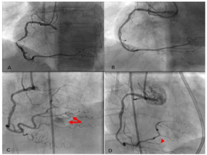
We described how we approached a grade II (Ellis classification), wire-related perforation in a small Posterolateral Branch (PLB) of the Right Coronary Artery (RCA) after an elective PCI was performed [Figure A to C]. The patient was initially treated conservatively, but returned to the catheterization suite after developing hypotension and breathlessness secondary to tamponade. A total of 150 mL of blood was drained with rapid hemodynamic improvement. A pigtail catheter was left in the pericardial space. The angiogram showed staining of contrast from the perforated artery that appeared unchanged from the index angiogram. We decided to perform no further interventions given the small size of the artery and the lack of visible extravasation of contrast.
The patient was brought back to the catheterization suite due to high pericardial output over a two-hour period. We decided to perform selective autologous fat embolization to achieve hemostasis after 20 minutes of balloon occlusion of the culprit vessel failed to do so. Fat particles were extracted with a mosquito clamp from a 10 mm-length skin incision over the inguinal area. The fat particles were 3 to 5 mm in size and were mixed with 5 mL of normal saline. We then used a 2.5F Transit micro catheter (Codman & Shurtleff, Inc. Raynham, MA USA) to test the amount of saline needed to flush a total of 0.5 mL of fat to the tip of the micro catheter. After two test injections on the sterile procedure table, we determined that 3 mL was needed to flush the fat particles. We then selectively engaged the culprit artery with the micro catheter, and injected the saline, fat mixture into the vessel. Angiography and ST segment elevation confirmed occlusion of the culprit vessel [Figure D/Video]. The pigtail catheter was removed 24 hours later, there was no residual effusion on the transthoracic echocardiogram. We want to emphasize that this technique is ideal for perforation of small arteries where coil embolization or balloon occlusion is not possible.
- Shimony A, Joseph L, Mottillo S and Eisenberg MJ. Coronary Artery Perforation During Percutaneous Coronary Intervention: A Systematic Review and Meta-analysis. Can J Cardiol. 2011; 27: 843-850. https://goo.gl/xRovXq
- He YL, Han JL, Guo LJ, Zhang FC, Cui M, and Gao W. Effect of Trans catheter Embolization by Autologous Fat Particles in the Treatment of Coronary Artery Perforation During Percutaneous Coronary Intervention. Chin Med J (Engl). 2015; 128: 745–749. https://goo.gl/sMMZLj


Sign up for Article Alerts