Infective Endocarditis: a Disease with Many Faces?
Bindi Patel and Ankit Agrawal*
Division of Cardiology, Department of Medicine, Montefiore Medical Center, Bronx, NY
*Address for Correspondence: Ankit Agrawal, Rutgers Robert Wood Johnson Medical School- Saint Peter’s University Hospital, 254 Easton Ave, New Brunswick, New Jersey 08901, USA, Tel: +1-732-853-7570; ORCID ID: 0000-0001-7885-6667; E-mail: [email protected]; [email protected]
Submitted: 27 October 2018; Approved: 29 November 2018; Published: 03 December 2018
Citation this article: Patel B, Agrawal A. Infective Endocarditis: a Disease with Many Faces. Int J Clin Cardiol Res. 2018;2(3): 076-079.
Copyright: © 2018 Patel B, et al. This is an open access article distributed under the Creative Commons Attribution License, which permits unrestricted use, distribution, and reproduction in any medium, provided the original work is properly cited
Keywords: Infective endocarditis; Complications of infective endocarditis; Multi-system manifestation
Download Fulltext PDF
Introduction
Infective endocarditis is a serious condition of the heart endocardium and cardiac valves. It is caused by a variety of microorganisms ranging from Streptococci, Staphylococci, Rickettsia or Coxiella. It can cause fever, night sweats and other constitutional symptoms, cardiac as well as multiple organ system involvement. Treatment includes prolonged antimicrobial therapy and emergent (within 24 hours), urgent (within few days) or elective cardiac surgery (after 1-2 weeks of appropriate antibiotic therapy) when indicated. This case outlines a case of subacute infective endocarditis and the clinical clues that may have prevented an impending cerebrovascular event.
Case Description
A 66 year old Caucasian woman with a past medical history of hypertension, hyperlipidemia and chronic obstructive pulmonary disease presented to the hospital with complaints of fevers, intermittent chills, and bilateral flank pain for two weeks. It was associated with burning sensation during micturition but there was no hematuria or purulent discharge in the urine. No history of trauma, renal stones or valvular disease or any previous similar complaints was reported. On presentation, the patient was hypotensive with blood pressure of 76/ 50 mm Hg, heart rate of 80/minute, respiratory rate of 30/minute and temperature of 99.3°F. Comprehensive physical examination was pertinent for poor dental hygiene and some mild costovertebral angle tenderness bilaterally. No cardiac murmur or any extra cardiac manifestation of infective endocarditis like Janeway lesions or Osler’s node was appreciated. Laboratory values were as per table 1. Intravenous fluids were started and the blood pressure was stabilized without vasopressor support. Blood cultures and urine cultures were sent with the suspicion of sepsis secondary to possible urinary tract infection. She was initially empirically managed with intravenous Ceftriaxone but then switched to intravenous Vancomycin after gram-positive bacteremia was reported on blood cultures.
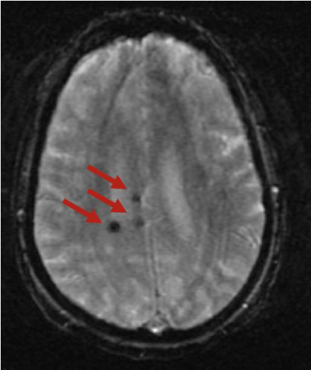
Urinary infections with gram positive organism is uncommon and this finding prompted us to suspect and further evaluate the heart for infective endocarditis by echocardiography. While awaiting blood culture sensitivity and echocardiography results, the patient experienced a sudden onset episode of left sided numbness and tingling, and thus the Rapid Response Team was called. On neurological assessment, patient had a diffuse decrease in sensation in left upper and lower extremities. Power was decreased and hyporeflexia was noted on the left side of the body. Cranial nerves and fundoscopic examination were within normal limits and cerebellar function was intact. Code Stroke was announced and immediate CT scan of head was obtained, which showed a small right parietal periventricular hyper density which was further evaluated by MRI with and without gadolinium contrast. The latter showed evidence of multi-territorial cerebral septic emboli (Figure 1). Echocardiogram showed a large mobile mass adherent to the posterior leaflet on the mitral valve in the left atrium that was highly suspicious for vegetation (Figure 2) and blood culture sensitivities revealed Streptococcus gordonii infection.
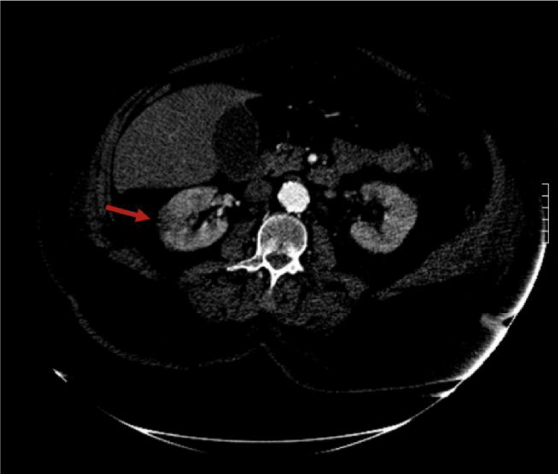
CT angiography of abdomen and pelvis was obtained which showed evidence of right renal infarct (Figure 3). This patient was medically managed in the intensive care unit with IV antibiotics, IV fluids and close hemodynamic monitoring. There were no signs and symptoms suggestive of heart failure. She was subsequently transferred to a tertiary care centre for mitral valve replacement due to massive valve destruction and is doing well on follow-up appointment.
Discussion
Subacute Infective Endocarditis (IE) has a gradually progressive course that causes cardiac damage slowly. Unfortunately, the presenting symptoms of this disease are heterogeneous in nature, and may be as nonspecific as musculoskeletal pain, fevers, and cardiac murmur, but can lead to major embolic event, such as renal infarcts, embolic strokes, and intracranial hemorrhage, as demonstrated in the case above [1,2]. The diagnosis is made by using modified Duke’s criteria (Table 2). Definite endocarditis is defined by documentation of 2 major criteria or 1 major and 3 minor criteria or 5 minor criteria. Common clinical features of IE include heart murmur (80-85%), fevers ≥ 100.4°F (80-90%), chills (40-75%), anorexia and malaise (25-50%), myalgias (15-30%), peripheral manifestations such as Osler’s nodes and Janeway lesions (2-15%), and neurological manifestations (20-40%) including stroke or transient ischemic attack, cerebral hemorrhage, mycotic aneurysm, meningitis, cerebral abscess, or encephalopathy [1,3]. Acute ischemic stroke is the most common neurological complication of IE, and the frequency of stroke is 8 per 1000 patient-days during the week prior to diagnosis [1,4,5]. Neurosurgical intervention may be required for certain patients with intracranial mycotic aneurysms that do not disappear after antimicrobial therapy or for aneurysms that enlarge or bleed. There are certain indications of early surgery in infective endocarditis which include heart failure, uncontrolled infections (defined as abscess, fistula or pseudoaneurysm formation) and for prevention of embolization (for vegetation >10 mm with previous history of embolization or associated with other surgical indications or vegetation >15 mm with accessible valve repair). Emergent surgery is required for heart failure with refractory pulmonary edema or hemodynamic instability. Enlarging vegetations, persistent fever or positive blood cultures even after 7-10 days of appropriate antibiotic therapy are other class I indications for urgent cardiac surgery.
Anterior mitral valve leaflet endocarditis confers the highest risk of stroke [4]. Streptococcus gordonii is part of the normal flora of the mouth and belongs to the viridans streptococci group, which are able to synthesize dextran from glucose, allowing them to adhere to fibrin-platelet aggregates at damaged heart valves after seeding into the bloodstream with minor oral traumas [6]. There are well defined high risk cardiac lesions which require prophylaxis before dental procedures like manipulation of gingival tissue, or the periapical region of the teeth or perforation of the oral mucosa and respiratory tract surgery. These risk factors include prosthetic heart valves, prior history of endocarditis, unrepaired cyanotic congenital heart disease, including palliative shunts or conduits, completely repaired congenital heart defects during the 6 months after repair, incompletely repaired congenital heart disease with residual defects adjacent to prosthetic material and post cardiac transplantation valvulopathy. This case demonstrates the importance of understanding subtle and non-specific clinical clues of infective endocarditis. Timely recognition may prevent multisystem involvement like cerebrovascular and renal complications. A wide spectrum of a disease presentation should always be considered while making a differential diagnosis which is a key to early recognition and optimal management.
Acknowledgement
This clinical case report was never presented anywhere else. No one else has contributed in any way in the process of preparing this case. There are no financial disclosures to mention.
- Kasper D, Fauci A, Hauser S, Longo D, Jameson J, Loscalzo J et al. Harrison's principles of internal medicine. 19th ed.
- Mourad O, Palda V, Detsky AS. A comprehensive evidence-based approach to fever of unknown origin. Arch Intern Med. 2003; 163: 545. https://goo.gl/MKTCvL
- Murdoch DR, Corey GR, Hoen B, Miró JM, Fowler VG, Bayer AS, et al. Clinical presentation, etiology, and outcome of infective endocarditis in the 21st century: the International Collaboration on Endocarditis-Prospective Cohort Study. Arch Intern Med. 2009; 169: 463. https://goo.gl/EQAuPk
- Morris NA, Matiello M, Lyons JL, Samuels MA. Neurologic complications in infective endocarditis: identification, management, and impact on cardiac surgery. Neurohospitalist. 2014; 4: 213-222. https://goo.gl/Wto8mZ
- Derex L, Bonnefoy E, Delahaye F. Impact of stroke on therapeutic decision making in infective endocarditis. J Neurol. 2009; 257: 315-321. https://goo.gl/uBffbw
- Scheld WM, Valone JA, Sande MA. Bacterial adherence in the pathogenesis of endocarditis. Interaction of bacterial dextran, platelets, and fibrin. J Clin Invest. 1978; 61: 1394-1404. https://goo.gl/ZNB9EZ


Sign up for Article Alerts