James Choueiry* , Gilbert Maroun, Charbel Kosaify, Mark A. Ghaida, Luke Heinrichs, Edgard Traboulsi, Davide Aliani and Charbel Maroun
James Choueiry1*, Gilbert Maroun2, Charbel Kosaify3, Mark A. Ghaida4, Luke Heinrichs4, Edgard Traboulsi5, Davide Aliani6 and Charbel Maroun4
1Cliniques Universitaires Saint-Luc, Brussels, Belgium
2American University of Beirut, Beirut, Lebanon
3Cliniques Universitaires Saint-Luc, Brussels, Belgium
4University of Alberta, Edmonton, Canada
5Central Military Hospital, Beirut, Lebanon
6DUniversity of Parma, Parma, Italy
*Address for Correspondence: James Choueiry, Cliniques Universitaires Saint-Luc, Brussels, Belgium, Tel: +003-247-679-5435; E-mail: jameschoueiry@hotmail.com
Dates: Submitted: 19 January 2018; Approved: 26 February 2018; Published: 28 February 2018
Citation this article: Choueiry J, Maroun G, Kosaify C, Ghaida MA, Heinrichs L, et al. Primary Hydatidosis in the Tibia, an Essential Preoperative Diagnosis. Int J Ortho Res Ther. 2018;2(1): 001-004.
Copyright: © 2018 Choueiry J, et al. This is an open access article distributed under the Creative Commons Attribution License, which permits unrestricted use, distribution, and reproduction in any medium, provided the original work is properly cited.
Keywords: Primary Hydatid Cyst; Bone; Knee Arthroplasty
Abstract
Primary hydatidosis of the tibia is a rare disease. In an endemic area, it should be considered in the differential diagnosis of a hypolucent osteolytic lesion on x-ray. If not properly managed, anaphylactic shock may occur intraoperatively, as well as increased recurrence of the disease. This is a case report of a primary tibial hydatid cyst, treated first with curettage and phenolizaton, and then after recurrence, with total knee arthroplasty. We will review the literature of the diagnosis and the treatment of a tibial hydatid cyst.
Introduction
Hydatidosis consists mainly of either cystic or alveolar hydatid diseases, they are caused by echinococcus granulosus and echinococcus multilocularis, respectively. Echinococcus granulosus is a tapeworm cestode and is acquired through a cycle in which the humans are intermediate hosts. Most patients have hydatid cysts of their liver, spleen, and lungs, which are the major filtering organs of these parasites. It is rare for the hydatid cyst to escape these filtering mechanisms and be present primarily in the tibia. When present in the tibia, it is difficult to diagnose radiologically. Therefore, there should be a high level of suspicion in endemic areas, such as mediterranean region, Central Asia, East Africa, Australia and New Zealand [1]. Suspicion of a hydatid cyst may prevent recurrence and complications. We studied a 33 year - old patient with primary hydatidosis in the tibia, in whom recurrence after curettage led to treatment with a total knee arthroplasty.
Patients and Methods
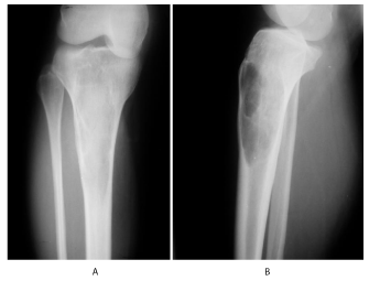
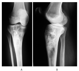
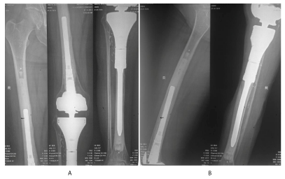
A 33 year - old female patient presented for right leg pain of a five week duration, exacerbated by weight bearing without disappearing at rest or at night. She estimates the pain as 6 over 10, relieved with non-steroidal anti - inflammatory agents. She is previously healthy without any history of trauma. There is mild edema, no redness, and mild increase of the pain upon palpation. There are no neurovascular abnormalities. Examination of the back, hip, and knee is normal. X-ray showed a large hypolucent spherical lesion in the proximal third of the tibia (Figure 1). MRI on T1 showed the lesion to be hypointense, and on T2 to be hyperintense (Figure 2,3). The lesion showed to be osteolytic, extending anteroposteriorly through the cortex at some levels, but it did not reach the articular surface, nor the surrounding soft tissues. Decision was taken to biopsy it. The pathology report showed lamellated cysts and scattered scoleces. Albendazole 400 mg was given orally twice per day for 2 months. Extensive curettage was done through excision of bone layers with a burr, then phenolization and grafting of cancellous bone chips inside the cavity. The patient was free clinically and radiologically of the disease for two years, then in the third year, multilocular cysts reappeared on x-ray (Figure 4), with possible extension to the articular surface. A decision of total knee arthroplasty was taken after wide resection of the diseased segment (Figure 5). Negative margins were obtained. The patient post operatively was rehabilitated; pain subsided with no radiological signs of recurrence. She was given albendazole 400 mg orally twice per day for 3 months.
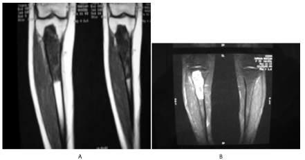 Figure 2: On the left: T1 coronal cuts showing hypointense proximal tibia lesion not reaching the articular surface, nor invading the surrounding soft tissues. On the right: T2 coronal cuts showing hyperintense lesion.
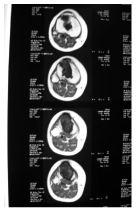 Figure 3: Transverse T1 cuts, showing the anteroposterior involvement of the tibia. No soft tissue invasion.
Discussion
The bones are infected in 0.45% to 2.5% of all cases of hydatid cysts [2], 10 % of bony involvement is in the tibia, 35% in the spine, 21% in the pelvis, 16% in the femur, 6% in the ribs, 4% in the skull, 4% in the scapula, 2% in the humerus, and 2% in the fibula [3]. The cysts are present in metaphyseal and epiphyseal areas first. The cortex is a hard structure, and the cysts have a thinner envelope due to the absence of a pericyst in the bones. Both of these factors will orient the development of the cysts into the area of least resistance towards the diaphysis, lysing the trabeculae, and slowly eroding into the cortex. As a result, the cysts grow in an irregular, branching fashion in the bone. It may take 20 years before the symptoms appear [4], making bone involvement less common in the younger population. If soft tissue is reached, the cyst will grow more rapidly and uniformly to form a large, spherical cyst. The patient may present with leg pain along with a palpable tumor, a pathologic fracture, or an infection. X-rays will show a single or multiple osteolytic cystic images [5], it can be multilocular with reactive sclerosis forming a honeycombing pattern [6]. Periosteal thickening may appear on x-ray if there is a pathological fracture [7]. The differential diagnosis is benign bone cyst, giant cell tumor, brown tumor in hyperparathyroidism, fibrous dysplasia, chondromyxoid fibroma, tuberculosis, metastasis, and aneurysmal bone cyst. In front of such images further investigation is required. CT scan will better evaluate, as well as the MRI, the lesion and its possible soft tissue invasion. CT scan will not show a calcified cystic rim. The latter only surrounds soft tissue cysts, not cysts in the bone [8,9]. It is suggested that calcification varies according to the disease course and duration since calcification is caused by dystrophic changes of dead cysts [3]. MRI will show hypointense on T1 and hyperintense on T2 daughter cysts separated by various amounts of fluid collections. MRI may show on T2 a low signal intensity if nonviable scolices are present in the daughter cysts upon the pathology [10]. A water-lily sign has been suggested as a pathognomonic MRI sign, it is the image of detachment of the germinative layer of the cyst from the pericyst, but it is rarely observed [11, 1]. It has been suggested that x-ray and even CT scan of hydatid disease are similar to those of metastases, giant cell tumors, tuberculosis, and bone cysts. Only MRI may aid in the diagnosis of hydatid cyst [12,13].
Serological tests depend on the state of the cysts. If the cyst is calcified and dead, immunoreactivity is low, but it increases if there are cyst rupture and spillage of the content. Common ways for the detection of anti-echinococcus IgG antibodies are the primary serology tests: the indirect hemagglutination test and the enzyme-linked immunosorbent assay [14].
There is still no more definite diagnostic test than the histopathological testing through bone biopsy [9]. The incision is anteromedial to avoid compartmental dissemination. Bone is biopsied taking care not to spill the dangerous highly anaphylactic cystic fluid. Desensitization to this fluid has been reported as ineffective [2]. Pathology reports show lamellated acellular cysts with germinal layers and scolices.
The main methods of treatment are suggested: curettage or radical resection, both with or without chemotherapy. Many have suggested the better results of a radical resection with or without pre - and post-operative chemotherapy [15-17]. They consider hydatid cysts as low grade malignancies with locally invasive properties. But Booz revealed good results with curettage and bone grafting with pre- and post-operative chemotherapy. He approved radical resection for “uncontrollable cases” [18] and considered total resection as the best solution in the fibula and ribs. After curettage, scolecidal agents may be used, such as formalin and hypertonic saline, but it has been suggested that they do not kill all microscopic daughter cysts [6]. Yildiz used PMMA to fill the cavity instead of bone grafts, because recurrence can happen in these bone grafts [6]. It was suggested that the thermal energy and free radicals released during polymerization have a scolicidal effect. Furthermore, monomers released from the PMMA are also toxic to living cells [19-21]. The chemotherapy consists of albendazole, mebendazole and praziquentel. Albendazole is usually preferred because it has the highest concentration inside the cysts.
If the patient lives in an endemic area or is in close contact with cattle, hydatid disease should be suspected. While in the bones it does not probably affect long term survival, it is difficult to cure. Furthermore, in the spine, it may result in paraplegia and high morbidity [22]. To break the cycle of transmission, in Bulgaria, infected sheep were killed in Karaguiosov, in 1970. In Kuwait, inspectors examine dog meat in the abbatoirs [18]. Many countries are considered endemic, but still no sufficient measures are taken to prevent this disease that is difficult to cure. Despite all radiological and serological tests, good history taking, searching for risk factors such as exposure to cattle or living in a rural area, is still the mainstay of suspicion of the hydatid cyst.
References
- Vasilevska V, Zafirovski G, Kirjas N, Janevska V, Samardziski M, Kostadinova-Kunovska S, et al. Imaging diagnosis of musculoskeletal hydatid disease. Prilozi. 2007; 28: 199-209. https://goo.gl/4Zb8kz
- Sapkas GS, Stathakopoulos DP, Babis GC, Tsarouchas JK. Hydatid disease of bones and joints. 8 cases followed for 4-16 years. Acta Orthop Scand. 1998; 69: 89-94. https://goo.gl/qhjeQG
- Merkle EM, Schulte M, Vogel J, Tomczak R, Rieber A, Kern P, et al. Musculoskeletal involvement in cystic echinococcosis: Report of eight cases and review of the literature. Am J Roentgenol. 1997; 168: 1531-1534. https://goo.gl/j6itjh
- Markakis P, Markakis S, Prevedorou D, Bouropoulou V. Echinococcosis of bone: clinico-laboratory findings and differential diagnostic problems. Arch Anat Cytol Pathol. 1990; 38: 92-94. https://goo.gl/8Atzkw
- Dahniya MH, Hanna MR, Ashebu S, Muhtaseb SA, El-Beltagi A, Badr S, et al. The imaging appearance of hydatid disease at some unusual sites. British Journal of Radiology. 2001; 74: 283-289. https://goo.gl/6mGPVP
- Yildiz Y, Bayrakci K, Altay M, Saglik Y. The use of polymethylmethacrylate in the management of hydatid disease of bone. J Bone Joint Surg Br. 2001; 83:1005-1008. https://goo.gl/1WgpVb
- Ersin Kuyucua, Mehmet Erdil b, Ali Dulgeroglua, Figen Kocyigitc, Arslan Bora. An unusual cause of knee pain in a young patient; hydatid disease of femur. Int J Surg Case Rep. 2012; 3: 403-406. https://goo.gl/mSwM18
- Booz MY. The value of plain film findings in hydatid disease of bone. Clin Radiol. 1993; 47: 265-268. https://goo.gl/Rh1fhi
- Torricelli P, Martinelli C, Biagini R, Ruggieri P, De Cristofaro R. Radiographic and computed tomographic findings in hydatid disease of bone. Skeletal Radiol. 1990; 19: 435-439. https://goo.gl/XQ5Y7U
- Garcia-Diez, Ros Mendosa LH, Villacampa VM, Cozar M, Fuertes MI. MRI evaluation of soft tissue hydatid disease. Eur Radiol. 2000; 10: 462-466. https://goo.gl/zZiozY
- Comert RB, Aydingoz U, Ucaner A, Arikan M. Water-lily sign on MR imaging of primary intramuscular hydatidosis of sartrius muscle. Skeletal Radiol. 2003; 32: 420-423. https://goo.gl/nMx7Ae
- Loudiye H, Aktaou S, Hassikou H, El-Bardouni A, El-Manouar M, Fizazi M, et al. 2003. Hydatid disease of bone: review of 11 cases. Joint Bone Spine. 70: 352-355. https://goo.gl/bmSP57
- Schneppenheim M, Jerosch J. Echinococcosis granulosus/cysticus of the tibia. Arch Orthop Trauma Surg. 2003; 123: 107-111. https://goo.gl/cQyXJ4
- Brunetti E, Kern P, Vuitton DA. Expert consensus for the diagnosis and treatment of cystic and alveolar echinococcosis in humans. Acta Trop. 2010; 114: 1-16. https://goo.gl/Lcs5SF
- Natarajan MV, Kumar AK, Sivaseelam A, Iyakutty P, Raja M, Rajagopal TS. Using a custom mega prosthesis to treat hydatidosis of bone: a report of 3 cases. J Orthop Surg (Hong Kong). 2002; 10: 203-205. https://goo.gl/yHtajZ
- Zlitni M, Ezzaouia K, Lebib H, Karray M, Kooli M, Mestiri M. Hydatid cyst of the bone: diagnosis and treatment. World J Surg. 2001; 25: 75-82. https://goo.gl/WJpA1i
- Mnaymneh W, Yacoubian V, Bikhazi K. Hydatidosis of the pelvic girdle-treatment by partial pelvectomy. A case report. J Bone Joint Surg Am. 1977; 59: 538-540. https://goo.gl/xs4BLF
- Booz MK. The management of hydatid disease of bone and joint. Journal of Bone and Joint Surgery. 1972; 54: 698-709. https://goo.gl/KKLaaV
- Strube HD, Komitowski D. Experimental studies of the treatment of malignant tumors with bone cement. In: Enneking WF, ed. Limb salvage in musculoskeletal oncology. New York: Churchill Livingstone. 1987: 459-469. https://goo.gl/biSVeL
- Mjoberg B, Pettersson H, Rosenqvist R, Rydholm A. Bone cement, thermal injury and the radiolucent zone. Acta Orthop Scand. 1984; 55: 597-600. https://goo.gl/CnEsL2
- Rock MG. Treatment of bone cysts and giant cell tumors. Current opinion in Orthopaedics. 1990; 1: 423-434.
- Yasir N Gashi, Walaa M Makki, Omiama AO, Ahmed M El Hassan. Paraplegia due to a large solitary hydatid cyst of the lumbar spine: a case report and review of the literature. Sudan Med J. 2013; 49. https://goo.gl/xZB55v
Authors submit all Proposals and manuscripts via Electronic Form!


