Case Report
A Rare Case of Primary Non-Hodgkin’s Lymphoma of The Bone in a Female Patient Addmited To Dubrovnik Hospital in 2013
Bekic M1*, Mikolaucic M1, Kosovic V2, Golubovic M1, Bilic V3 and Lalovac M4
1Department of Orthopaedic and Traumatollogy, County Hospital Dubrovnik, Dubrovnik, Croatia
2Department of Radiology, County Hospital Dubrovnik, Dubrovnik, Croatia
3Clinical hospital center Sisters of Charity, Clinic of traumatology, Zagreb, Croatia
4Clinical hospital Merkur, Clinic for internal medicine, Zagreb, Croatia
*Address for Correspondence: Dr. Marijo Bekic, Department of Orthopaedic and Traumatollogy, County Hospital Dubrovnik, Dubrovnik, Croatia, Tel: +385 20431858; E-mail: marijob@bolnica-du.hr
Dates: Submitted: 26 August 2017; Approved: 28 August 2017; Published: 05 September 2017
Citation this article: Bekic M, Mikolaucic M, Kosovic V, Golubovic M, Bilic V, Lalovac M. A Rare Case of Primary Non-Hodgkin’s Lymphoma of The Bone in a Female Patient Addmited To Dubrovnik Hospital in 2013. Int J Ortho Res Ther. 2017;1(1): 016-020.
Copyright: © 2017 Bekic M, et al. This is an open access article distributed under the Creative Commons Attribution License, which permits unrestricted use, distribution, and reproduction in any medium, provided the original work is properly cited.
Keywords: Primary Non-Hodgkin’s Lymphoma of Bone; Pathological Fracture; Chemotherapy
Abstract
Primary Non-Hodgkin’s Lymphoma of The Bone (PLB) is rare entity [1,2,3]. Patients generaly present with localised bone pain, soft-tissue swelling or palpable mass. Pathological fracture of the proximal femur and humerus secondary to soft-tissue tumors is well documented in the literature. Lymphomas presenting primarly at these sites with pathological fracture is unusual. A review of the world literature shows that the incidence of the skeletal manifestations from NHL is less than 5%, and in all these cases, bony involvement was reported many years after presentation of the primary cancer. Histopathologically, PLB usually represents diffuse large B-cell lymphoma. We report 56-year-old female patient case report of Primary non-Hodgkin’s limphoma of proximal femur and proximal femur with pathological fracture and management. In january 2014. A 56-year-old woman was diagnosed with stage IV B primatry large-cell diffuse primary non-Hodgik’s lymphoma. After one year of initial diagnostic procedures and chemotherapy with rituximab together with cyclophosphamide, vincristine, procarbazine, and prednisolone she achieved a total response.
Introduction
Primary Lymphoma of The Bone (PLB) is a rare extranodal presentation of Non-Hodgkin’s Lymphoma (NHL) Jacobs Kurosawa. It was first described by Oberling in 1928 [4,5]. It accounts for approximattely 3% of the malignant bone neoplasms and is comprised of less than 5% of of all extranodal non-Hodgkin’s lymphomas. An osseous involvement of a lymphoma is generally seen as a part of a multi-system disemination. Primary lymphoma of the bone can be defined as a lymphoma which occurs in the bone without any evidence of a distal nodal or an extra-nodal tissue involvement [5,6]. PBL can involve any part of the sceleton, but a trend exists in favour of the bones with persistent bone marrow. The femur is the most common site and it is affected in 29% of the cases. The other sites inculde the pelvis, humerus, head and neck, and the tibia [4-7]. The clinicall presentation depends upon the rate of the tumor cell proliferation and the initial localisation. The patients generally present with localized bone pain, and less frequently, with a soft-tissue swelling or a palpable mass. On conventional radiology, PLB has a widely variable imaging manifestations which consists of either „lytic destructive pattern“ or a „blastic sclerotic pattern“ [8]. Pathological fractures may be present in approximately one quarter of the cases, as were seen in our patients.
Diagnostic testing typically include: Complete Blood Cell Count (CBC); peripheral blood smear; routine serum chemistries; serum Lactate Dehydrogenase (LDH) level; bone marrow aspirate and biopsy; bone biopsy; chest radiography; plain films; bone scan; Computed Tomography (CT) scanning; Magnetic Resonance Imaging (MRI). When detectable, primary NHL of bone can have a heterogeneous appearance with lytic, blastic, and mixed lesions reported [6,9,10]. Additional imaging features include periosteal reaction, soft-tissue extension,, pathologic fracture, and cord compression [6]. The radiologic differential diagnosis includes benign (reactive conditions, osteomyelitis)and malignant entities (Hodgkin lymphoma, sarcoma, neuroblastoma, metastatic disease).
Materials and Methods
A 56-year old female patinet presented in December 2013. with pain in the chest, thoracal spine, epigastric and right scapular region for one month duration. Clinically, the patient had history of 3 kg weight loss and fatigue. On examination, she was found to be hypertensive (160/90 mmHg) and had no palpable hepatosplenomegaly or lymphadenopathy. In past the patient had arterial hipertension, she takes no therapy. In 2009. The patient underwent gastoenterollogyst examination : Ezophagogastroscopy showed an moderate gastroesophageal reflux into the distal oesophageos, with hiperemic, vulnerable mucose. Diagnosis: Sy Gerd gr 2. EKG : QS in V1-V3.
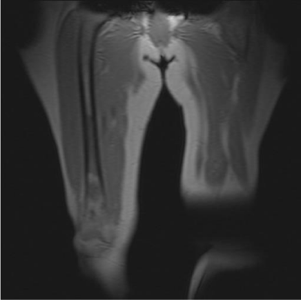
Antihipertensive and analgetic therapy was oredered and the difficulties have been reducted. After twenty days the patient was examined by Neurologyst because of the sudden pain in the thoracal spine which spreaded into the sternal region and in the right leg as well. She uses anti-pain drugs, but with no improwement. Neurologicaly, there were signs of hipoaesthaesia in the L4, L5 dermatoma on the right. Lasegue test terminally positive [Figure 1].
 Figure 1: Coronal T1-weighted image reveals few low signal lesions in the marrow of the mid and distal right femoral shaft which contrasts with areas of high signal intensity of normal marrow fat.
The MR of the thoracal and LS spine was recomended.
Leukocites: 4,80. (Ref. 3,40-9,70); Haemoglobine (g/L): 135, (Ref. 119-157), Trombocites(10e9/L): 227 (Ref. 6,80-10,40): CRP (mg/L) 14,48 (0,00-5,00), Alcal-phosphathase (U/L) 124 (64-153): Troponine (ng/ml) > 0,01 (Ref. 0,000-0,100): Amilase (U/L) 345 (Ref. <400).
X Ray of the thoracal spine : less calcifisated body and archs of thoracal spine, with subchondral osteosclerosis surface plate with signs of partial degenerative spondilosis. The height of the thoracal spine corps was normal. Intervertebral space between TH 7 – 8 was slightly reduced.
The chest radiograph and the ultrasound of the abdomen were normal as was her full blood count and biochemical prophiles.
Ginaecological exmination , esophagogastroendoscopy and colonoscopy, MMg, oftalmological, dermatological examination were negative.
The patient was immobilisaed with Jewett orthosis.
One month after the initial examination by Internal medicine specialist and two months of the duration of pain, the patient was reported to our Orthopedic and traumatology department with a pathological fracture of the TH 5 and L 2 spine corps, proved with MRI diagnostics.
Ca (mmol/L) 2,39, Ca-dU (mmol/L) 6,1.
Computorised tomography (CT) scan of the right femur showed small lytic destructions of bone corticalis 10 cm of lenght in the mid-shaft of the right femur. There were no signs of bone compacta destruction or bone expansion. Pathologic supstrate which infiltrated medularry narow was also presented. Acording the radiomorphologicall characteristics, diferential diagnosticly the femoral lesion points on low-grade chondrosarcoma.
MRI findings revealed a large expandible lytic infiltrative lesion of the medullary canale with very discrete diffuse bone compacta destruction at the level of the mid and distal shaft of the right femur, for 16 cm of craniocaudale diamater was recorded. There was absence of bone corticalis lytic lesion. There were no pathologicall findings of extraosseal soft tissue or periosteal reaction. Radiomorphologicall caracteristic were suggestive of metastasis or an osteogenic tumor.
The first biopsy of the veretbral body showed no signs of tumor tissue. The new intramedular biopsy of the right femur was performed after seven days: the seven samples were positive on vimentine and LCA, negative on pancreatokeratine, S-100 and CD 3. Histologicaly examination showed diffuse large B-cell NHL (high gradus).
The biopsy report was suggestive of a lymphoproliferative disorder which involved the bone.
The tissue was sent for a histopathological examination. The final pathological diagnosis was a primary lymphoma-NHL, diffuse large B-cell type (non-GC subtipe) of the shaft of the femur.
Imunohystochemically the cells were positive for CD20+, BCL6+, BCL2+,CD10 focal+(less than 30% cells), MUM1+, CD5-, CD3-, EBV-LMP1-. Clinical stady IV E [Figure 2].
 Figure 2: Sagittal T1 –weighted image reveals few low signal lesions in the marrow of the mid and distal femoral shaft without extraosseal soft tissue mass or periostal reaction.
The patient recived chemotherapy which consisted cyclophosphamide, adriamycin, vincristine, and prednisone (CHOP).
Total body scintigraphy was performed with 99m-Tc MBq 740. After 12 months of the diagnosis, there has been no relapse either clinically or radiologically.
Two month after the initial examination, the patient suffered pain in the right femur. She denides pain at the spine. She was verticalised with Jewett orthosis and walker. The primary tumor site was not founded.
A three months after the final diagnosis, the treatment with chemothery is performed: 4+4 cyclus.
She achieved a partial response with rituximab together with cyclophosphamide, vincristine, procarbazine and prednisolone.
Three years after the first symptoms and initial examination the patient still suffered cervical and lumbal spine pain with rare cases of vertigo. She used cervical collar, Jewett orthosis and analgetics.
PET-CT didnt recognised any signs of malignant disease or metabolic activity. The conclusion: the remission of disease has been achieved [Figure 3].
Discussion
Most cases of primary lymphomas of the bone are the NHL type and the Diffuse Large B-Cell Lymphoma (DLBCL) subtype. Primary NHL of the bone (primary bone lymphoma) is a rare condition, accounting for less than 1-2 % of adult NHL, and less than 7-10 % primary bone tumors [1,2]. The majority of the cases are limited disease by the Ann-Arbour staging system and occurs in adults of age 45-50 and it shows a male preponderance with a male to female ratio of 3:2. [4, 8-13].
Primary Lymphoma Of Bone (PLB) accounts for 3% to 7% of primary neoplasms of bone and must be distinguished from more common bone tumors in the pediatric population such as osteosarcoma, Ewing sarcoma, and other small round blue cell tumors. In this study, pathology databases from 4 institutions were queried for PLB in individuals 1 to 21 years old [14].
The femur (29%) is the most common site, followed by the pelvis (19%), humerus (13%), skull (11%) and the tibia (10%) [15].
Some series have found that the long bones and the flat bones are equally affected [12].
The clinical presentation includes local pain, swelling and a pathological fracture. The diagnosis is established by biopsy. CT scan of the abdomen and the chest to assess the lymph node involvement and serum LDH estimation are done as a a part of the stagging procedure. [16].
The most common pathology subtype is diffuse large B-cell lymphoma [12,16].
A pathological fracture of the long bones, which are secondary to the soft-tissue tumors had been well documented in the literature: lymphomas which present primarily at these sites with pathological fractures are unusual.
The most common pattern „the lytic destructive pattern“ , presented as permeative, moth-eaten or focal lyses with well-defined margins is reported in 70% of the cases [8] [Figure 4].
 Figure 4: Axial T1-weighted image reveals some focal low intensity signals in distal femoral part in the affected side.
Pathological fracture in bone lymphoma is uncommon. In a large series of 131 patients with primary bone lymphoma over 22-year period, one third had lymphoma involvement of the long bones with pathological fracture occuring in nine patients [12]. In contrast, in a retrospective analysis of 36 patients with primary (n=17) and secondary (n=19) bone lymphoma surgically treated at an orthopaedic center over a 15-year period, pathological fracture of the proximal femur of humerus was observed in three patients only [5]. Our patient illustrated pathological fracture secondary to lymphoma involvement of the femor, which is uncomon even in patients with lymphoma involvement of the long bones.
In a study of sixteen patients with primary NHL in the vertebrae of the spine, who were treated between 1994 and 2006 (10) 5 of them had radicular pain. Our patient had also radicular pain, but no motor signs of neurological dephicite[18] [Figure 5 a,b].
 Figure 5a.b: Sagittal T2 FS (a) and coronal T2 FS (b) images show high signal intensity of the lesions in central and distal part of the femur and linear abnormalities within the cortical bone in the mid part of the femoral shaft
The overall 5-year survival rate is 49.6%, with a 10-year survival rate of 30.2%. Median overall survival is 4.9 years (95% CI: 3.9, 6.1). In multivariable analysis, age (p<0.0001), marital status (p=0.006), and appendicular vs. axial tumor location (p=0.004) is found to be independent predictors of survival [19].
Anaemia, elevated lactate dehydrogenase (LDH), Alcaline Phosphatase (ALP), Erythrocyte Sedimentation Rates (ESRs), platelet counts, and calcium levels have been reported with primary non-Hodgkin lymphoma of bone [6,11].
Our patients has no elevated laboratory tests values [Figure 6].
 Figure 6: STIR T2-weigted sagittal image shows high signal intensity of the lesions in central and distal part of the femur.
The treatmant for primary NHL of bone includes chemotherapy, the optimal sequence of treatmanet is CHOP-R 8Cyclophosphamide, doxorubicin, vincristine, prednisolone, rifuximab) regimen [4,11-13,17]. As necessary, stabilisation of a pathologic fracture will occur before the initiation of other therapies [6,12].
The patient recived chemotherapy which consisted cyclophosphamide, adriamycin, vincristine, and prednisone (CHOP). After 12 months of the diagnosis, there has been no relapse either clinically or radiologically [Figure 7]. Here presented case agreed with the literature on primary bone lymphoma, in which the diagnostic problem and an excellent prognosis of a malignant tumor have been emphasized [15].
References
- Rojas N, Fernandes C, Conde M, Montala N, Fornos X, Rossello L, et al. Non-Hodgkin's lymphoma and atypical neck pain: A case report. Reumatol Clin. 2017. https://goo.gl/av8NwM
- Wang J, Fan S, Liu J, Song B. A rare case report of primary bone lymphoma and a brief review of the literature. Onco Targets Ther. 2016. https://goo.gl/nVDZCx
- Hayase E, Kurosawa M, Suzuki H, Kasahara K, Yamakawa T, Yonezumi M, et al. Primary Bone Lymphoma: A Clinical Analysis of 17 Patients in a Single Institution. Acta Haematol. 2015. https://goo.gl/C3FDhi
- Barbieri E, Cammelli S, Mauro F, Perini F, Cazzola A, Neri S, et al. Primary non-Hodgkin's lymphoma of the bone: treatment and analysis of prognostic factors for Stage I and Stage II. Int J Radiat Oncol Biol Phys. 2004. https://goo.gl/eaoJH8
- Durr HR, Muller PE, Hiller E, Maier M, Baur A, Jansson V, et al. Maligannat lymphoma of bone. Arch Orthop Trauma Surg 2002. https://goo.gl/Qe286p
- Glotzbecker MP, Kersun LS, Choi JK, Wills BP, Schaffer AA, Dormans JP. Primary non-Hodgkin's lymphoma of the bone in children. J Bone Joint Surg Am. 2006. https://goo.gl/7c4CjL
- Hsieh PP, Tseng HH, Chang ST, Fu TY, Lu CL, Chuang SS. Primary non-Hidgkin's lymphoma of bone: a rare disorder with high frequency of T-cell phenotype in southern taiwan. Leuk Lymphoma. 2006. https://goo.gl/U83GCX
- Krishan A, Shirkoda A, Tehranzadeh J, Armin AR, Irwin R, Les K. Primary bone lymphoma: A radiographic-MR imaging correlation. Radiographics. 2003; 23: 1371-83. https://goo.gl/t9WKPK
- Rappaport DC, Chamberlain DW, Shepherd FA, Hutcheon MA. Lymphoproloferative disorders after lung transplantation: imaging feautres. Radiology. 1998; 206: 519-24. https://goo.gl/aZ857L
- Stein ME, Kuten A, Gez E, Rosenblatt KE, Drumea K, Ben-Schachar M, et al. Primary lymphoma of bone-a retrospective study. Experience at the Northern Israel Oncology Center (1979-2000). Oncology. 2003. https://goo.gl/LvA7Ui
- Lima FP, Bousquet M, Gomez-Brouchet A, de Palva GR, Amstalden EM, Soares FA, et al. Primary diffuse large B-cell lymphoma of bone displays preferential rearrangements of the c-MYC or BCL2 gene. Am J Clin Pathol. 2008. https://goo.gl/UV8UtM
- Peng X, Wan Y, Chen L et al. Primary non-Hodgkin lymphoma of the spine with neurologic compression treated by radiotherapy and chemoterapy alone combined with surgical decompression. Oncol Rep. 2009. https://goo.gl/vRVbFJ
- Power DG, McVey GP, Korpanty G, Treacy A, Dervan P, OKeane C, et al. Primary bone lymphoma: single institution case series. 2008. https://goo.gl/4bzhCT
- Ramadan KM, Shenkier T, Sehn LH et al. A clinicopathological retrospective study of 131 patients with primary bone lymphoma: A population-based study of successively treated cohorts from the British Columbia Cancer Agency. Ann Oncol. 2007. https://goo.gl/ETUfQ3
- Chisholm KM, Ohgami RS, Tan B, Hasserjian RP, Weinberg OK. Primary lymphoma of bone in the pediatric and young adult population. Hum Pathol. 2017. https://goo.gl/i18cn6
- Salter M, Sollacico RJ, Bernreuter WK, Weppelmann B. Primary lymphoma of the bone: The use of MRI in the pretreatment evalution. Am J Clin Oncol. 1989. https://goo.gl/pqWuYm
- Siddiqui YS, Khan AQ, Sherwani M. Pathological fracture in primary non-Hodgkin Lymphoma of the bone: A case series with review of the literature. L Clin Diagn Res. 2013. https://goo.gl/DcuKox
- Singh T, Satheesh CT, Lakshmaiah KC, Suresh TM, Babu GK, Lokanatha D, et al. Primary bone lymphoma: A report of two cases and the review of the literature. Journal of Cancer Research and Therapeutics. 2010. https://goo.gl/dSekyv
- Jacobs AJ, Michels R, Stein J, Levin AS. Socioeconomic and demographic factors contributing to outcomes in patients with primary lymphoma of bone. J Bone Oncol. 2015. https://goo.gl/4L5d6V
Authors submit all Proposals and manuscripts via Electronic Form!


