Rodrigo Liceaga-Reyes1*, Stefanny Romero-Oyuela2, Gladys Acuna-Gonzalez3 and Cheryl Chavez Santisbon4
1Oral and Maxillofacial Surgeon. Associate Professor. Specialties Hospital, Campeche, Mexico
2Maxillofacial Prothesis Trainee Program, UNAM, Mexico
3Oral and Maxillofacial Surgeon. Associate Professor. Specialties Hospital, Campeche, Mexico
4General Dentist, associated to Maxillofacial Surgery Department at Specialties Hospital, Campeche, Mexico
*Address for Correspondence: Rodrigo Liceaga-Reyes, Hospital de Especialidades, Av Lazaro cardenas 208 Col Las Flores, CP 24097, San Francisco de Campeche, Campeche, Mexico, Tel: (52) 5585786138; E-mail: [email protected]
Dates: 28 June 2017; Approved: 05 July 2017; Published: 07 July 2017
Citation this article: Liceaga-Reyes R, Romero-Oyuela S, Acuna-Gonzalez G, Santisbon CC. Aggressive Behavior of Antralpseudocyst in the Maxilla: Case Report. Sci J Res Dentistry. 2017;1(1): 021-024.
Copyright: © Liceaga-Reyes R, et al. This is an open access article distributed under the Creative Commons Attribution License, which permits unrestricted use, distribution, and reproduction in any medium, provided the original work is properly cited.
Keywords: Antral pseudocyst; Benign neoplasia; Enucleation; Reconstruction
Abstract
The antralpseudocyst originates from the accumulation of serous inflammatory exudate in the sinus membrane without a specific etiology, this cyst has not of age group or gender preference. Radiographically, it is associated with a soft dome-shaped radiopaque pattern. This case is about a male patient of 58 years of age, with increased volume in the malar and left genic region of smooth, fluctuating consistency, which crackles at the pressure. Intraorally with the corresponding increase in volume in the sac, without changes in the oral mucosa. The tomography showed a radiolucent lesion that occupies and destroys left jaw and orbital floor. Thus, complete enucleation of the lesion and reconstruction of adjacent structures were performed. Clinical and imaging follow-up was carried out without postoperative complications and 8 years free of injury. It is of vital importance a correct diagnosis to guide the treatment adequately, however it is not necessary to underestimate the behavior of benign lesions and described as non-invasive.
Introduction
The antralpseudocyst is a rare pathology that originates from the accumulation of serous inflammatory exudate in the sinus membrane that may or may not produce sessile inflammation. No specific etiology is known but has been associated with viral infections or recurrent upper respiratory tract irritation [1].
Radiographically, it is associated with a soft, well delimited radiopaque pattern that is described as a dome-shape in the walls of the maxillary sinus mainly in the lower one [1,2,3]. Differential diagnoses must be taken into account both tumors and cysts Odontogenic and not odontogenic. Its diagnosis is made by histological study with material obtained from fine needle aspiration in which a yellowish viscous liquid is present and histologically presents a cylindrical pseudo-stratified epithelium with presence of inflammatory infiltrate and blood cells [4].
Case Report
This is a 58-year-old male patient with no relevant history for the condition, who reported having started some months ago with an increase in volume in the malar and left genic region, without receiving treatment (Figure 1). It does not mention suffering from systemic diseases or being under medical treatment. It denies a history of trauma or previous surgeries in the affected region. On physical examination he found his vital signs within normal parameters, with fascies characterized by a slight increase in volume in left side of the face, at palpation without local change in temperature, with a smooth, fluctuating consistency, which crackles at the pressure. Intraorally with the corresponding volume increase in the back of the sack, without changes of coloration in the oral mucosa, of adequate hydration, without liquid outlet or secretion. Incomplete permanent dentition with poor oral hygiene.
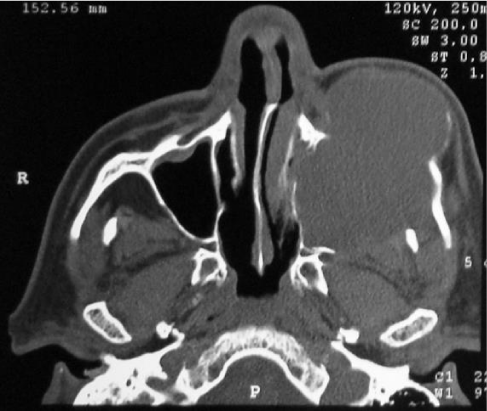
On aspiration biopsy was obtained by easily obtaining a light brown coloring liquid with blood residues. Under local anesthesia, incisional biopsy was performed for histopathological study, which later reported the antralpseudocyst, and its surgical enucleation was scheduled. Complementary imaging studies were carried out, which showed a radiolucent lesion in his tomographic image that occupies and destroys the left side of the jaw, including the floor of the orbit (Figure 2). A stereolithographic model was developed for the planning of the procedure (Figure 3).
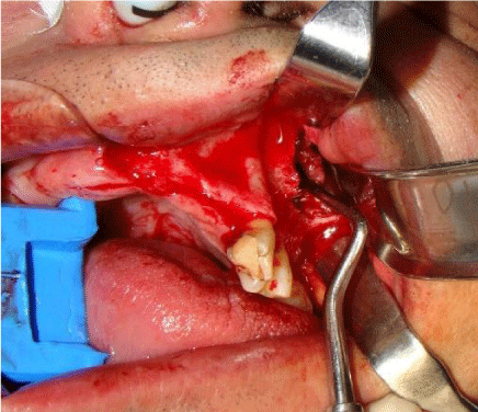
Subsequently, in the operating room under general anesthesia with orotracheal intubation, a crestal approach with extension throughout the entire alveolar process was performed intraorally to expose the anterior portion of the maxilla on the left side, allowing the complete enucleation of the lesion without the need for cutaneous incisions (Figure 4).
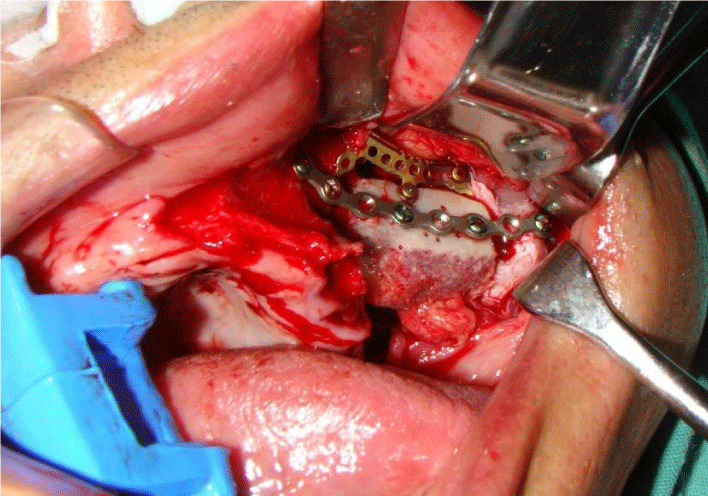
According to the planning carried out through the same approach was placed a mesh for orbit that was fixed to the bone remnant. Simultaneously with the enucleation, the anterior iliac autograft was taken to obtain a corticomedular block that was fixed with a system 2.0 osteosynthesis material (Figure 5).
The surgical specimen was submitted for histopathological examination; in its microscopic description, loose fibroconective tissue was identified, with ducts of dilated salivary glands, and in the interior myxoid aspect material without cover epithelium, establishing the diagnosis of antralpseudocyst (Figure 6).
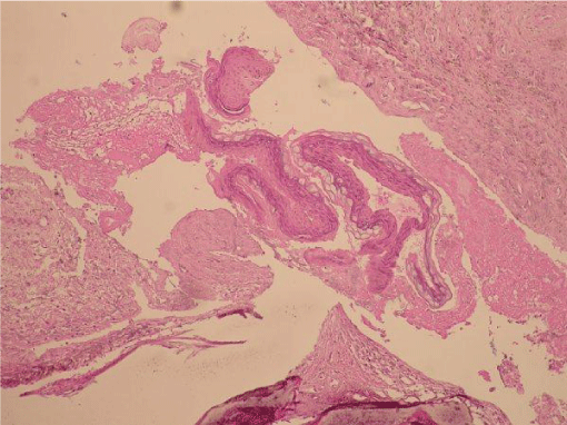 Figure 6: Histopathology of the case showing fibrous connective tissue and the absence of the epithelium.
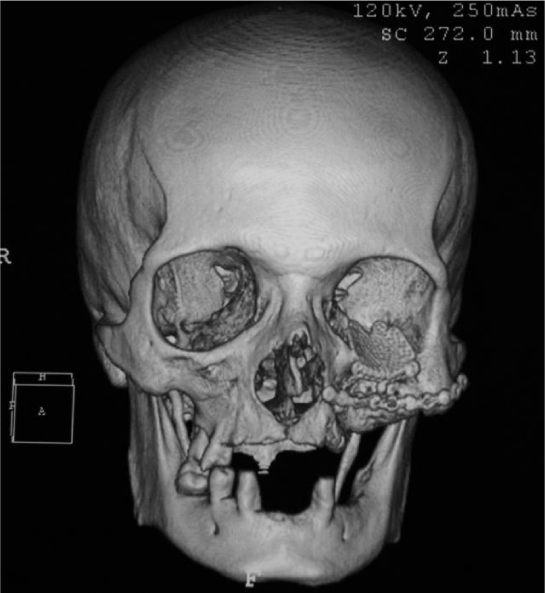
Clinical and imaging follow-up was carried out without complications, achieving adequate facial symmetry without compromising acuity or ocular mobility, and with complete intraoral cicatrization (Figure 7). The wound of the protruding area in the hip course without eventualities. At 8 years of the procedure there are no relapse data, with complete integration of the graft (Figure 8).
Discussion
AntralPseudocysts are generally found as a finding during the planning of surgical procedures, mostly implantological [5,6,7] involving maxillary sinus involvement because most of them do not present clinical or radiographic manifestations due to their size [8,9]. However in our case the patient had a significant volume increase that caused facial deformity, loss of the back of the sac and mild pain symptomatology.
It is important to perform a correct diagnosis of the antral pseudocyst because it usually does not require treatment for its size less and does not inflate adjacent tissues [10], in counter position to some of the differential diagnoses that require surgical treatment such as mucocele [11], as well as differentiate it of the majority of odontogenic cysts and tumors requiring enucleation or resections with injury-free margins [12]. In the present case the behavior of the cyst was completely different from that described in the literature as it infiltrated and caused destruction of the floor of the orbit and left jaw.
The aspiration of the antralpseudocyst is described as a light yellow liquid however in this case the obtained exudate had light brown coloration with blood remains. The aspiration of the lesion is not a treatment option because it is necessary to enucleate the tissue completely [4,5].
The most used surgical techniques as treatment are endoscopic surgery and open surgery Caldwell-Luc type [7,10,12]. In the present case it was necessary to perform a broader approach due to the size of the lesion and the involvement of adjacent structures, the usual behavior of antralpseudocyst, which is described as non-invasive, however, in this case reconstruction was necessary by a Titanium mesh for orbit that was fixed to the bone remnant and simultaneously to the enucleation the anterior iliac autograft was taken to obtain a corticomedular block that was fixed with system 2.0 osteosynthesis material.
Conclusion
It is vital to carry out the diagnosis of each of the entities that may present in the head and neck as this determines the success, the type of treatment and the alternatives available for each of the diagnoses.
Although the antralpseudocyst in most cases is found as a finding in routine imaging studies, when it is diagnosed, it is important to perform a complete removal of the lesion and perform a histopathological study.
In the case of the antralpseudocyst, it is important not to neglect the behavior of the lesion, since we have shown how it can invade and compromise the integrity of adjacent structures.
References
- SetteA, DrummondM, AlvesR. Differential diagnosis of antral pseudocyst. A case report, Baltic Dental and Maxillofacial Journal, 2013; 15: 93-5. https://goo.gl/tr7stH
- Baykul D, Fındık Y. Maxillary sinus perforation with presence of an antral pseudocyst, repaired with platelet rich fibrin. Ann MaxillofacSurg. 2014; 4: 205–207. https://goo.gl/Jrc4Nw
- Chiapasco M, PalomboD. Sinus grafting and simultaneous removal of large antral pseudocysts of the maxillary sinus with a micro-invasive intraoral access. Int. J. Oral Maxillofac. Surg. 2015; 44: 1499–1505. https://goo.gl/ByvfCY
- Aditya Tadinada, Elnaz Jalali, Wesam Al-Salman, Shantanu Jambhekar, Bina Katechia and Khalid Almas, et al. Prevalence of bony septa, antral pathology, and dimensions of the maxillary sinus from a sinus augmentation perspective: A retrospective cone-beam computed tomography study Imag Science in Dentistry 2016; 46: 109-15. https://goo.gl/ikXn9W
- Tang ZH, Wu MJ, Xu WH. Implants placed simultaneously with maxillary sinus floor augmentations in the presence of antral pseudocysts: a case report Int. J. Oral Maxillofac. Surg. 2011; 40: 998–1001. https://goo.gl/L2aSZq
- Lin Y, Hu X, Metzmacher AR, Luo H, Heberer S, Nelson K. Maxillary Sinus Augmentation Following Removal of a Maxillary Sinus Pseudocyst After a Shortened Healing Period. J Oral MaxillofacSurg. 2010; 68: 2856-2860. https://goo.gl/EBTN2e
- Kara I, Küçük D, PolatS. Experience of Maxillary Sinus Floor Augmentation in the Presence of Antral Pseudocysts, J Oral MaxillofacSurg, 2010; 68: 1646-1650. https://goo.gl/yUtbpk
- Mardinger O, Manor I, Mijiritsky E, Hirshberg A. Maxillary sinus augmentation in the presence of antral pseudocyst: a clinical approach. Oral Surg Oral Med Oral Pathol Oral RadiolEndod 2007; 103: 180-4. https://goo.gl/Ys4TVT
- Giotakis EI, Weber RK. Cysts of the maxillary sinus: a literature review. Int Forum Allergy Rhinol. 2013; 3: 766–71. https://goo.gl/rXrx1x
- Moon IJ, Kim SW, Han DH, Shin JM, Rhee CS, Lee CH, et al. Mucosal cysts in the paranasal sinuses: long-term follow-up and clinical implications. Am J Rhinol Allergy 2011; 25: 98–102. https://goo.gl/nQCGu5
- Wang JH, Jang YJ, Lee BJ. Natural course of retention cysts of the maxillary sinus: Long-term follow-up results. Laryngoscope 2007; 117-341. https://goo.gl/kGS51J
- Schalek P, Otruba L, Hornáčková Z, Hahn A. Mucosal maxillary cysts: long-term subjective outcomes after surgical treatment. Eur Arch Otorhinolaryngol. 2013; 270: 2263-6. https://goo.gl/hn1Unc
Authors submit all Proposals and manuscripts via Electronic Form!


