Danielle A. Cunningham, MS*and Robert Weidling, MS
University of Missouri-Kansas City School of Medicine, 2411 Holmes St, Kansas City, MO 64108
*Address for Correspondence: Danielle A. Cunningham, University of Missouri-Kansas City School of Medicine, 2411 Holmes St, Kansas City, MO 64108
Dates: Submitted: 13 December 2016; Approved: 31 January 2017; Published: 03 February 2017
Citation this article: Cunningham DA, Weidling R. Cerebral Abscesses Masquerading as Metastasis . Sci J Neuro Neurosur. 2017;3(1): 011-014
Copyright: © 2017 Cunningham DA, et al. This is an open access article distributed under the Creative Commons Attribution License, which permits unrestricted use, distribution, and reproduction in any medium, provided the original work is properly cited.
Keywords: Brain abscess; Dental procedure; Immunosuppression; Infectious disease; Crohn's disease; Viridans streptococci
Abstract
In a patient presenting with focal neurologic signs and multiple brain lesions, the differential diagnosis includes cerebral abscesses and metastatic disease. While the two conditions are commonly distinguished by the patient's history and neuroimaging, the diagnosis is challenging in select patients.
A 56-year-old gentleman with a history of Hepatitis C, Autoimmune Hepatitis, and Crohn's disease controlled on prednisone and infliximab presented with a recent seizure, left hemiparesis, and acute respiratory failure. Workup was significant for metabolic acidosis, a ring enhancing 22 x 20 x 17 mm mass in the frontal lobe on head CT, and a normal CT angiography of the chest. An MRI of the brain revealed a 28 x 22 x 26 mm mass with surrounding edema in the anterior superior lateral right frontal lobe and a smaller 11 x 11 x 10 mm ring enhancing lesion with surrounding edema in the right head of the caudate. The frontal abscess was incised and drained, and the cultures revealed Viridans streptococci. The patient was started on intravenous antibiotics, and he had complete return to respiratory and neurologic function. Once alert, the patient reported a long history of severe periodontal disease with recent deep dental cleanings, likely providing a nidus of infection. This case exemplifies the difficulty in discerning cerebral abscess from malignancy in select cases, and the risk of infection in patients on immunomodulating medications.
Abbreviations
BiPAP: Biphasic Positive Air Pressure; BP: Blood Pressure; CT: Computed Tomography; HR: Heart Rate; IV: Intravenous; MRI: Magnetic Resonance Imaging; PE: Pulmonary Embolism; RR: Respiratory Rate
Introduction
In the United States, approximately 1,500 to 2,500 cerebral abscesses are diagnosed each year [1]. This number is increasing, likely as a result of more patients taking immunomodulating medications [2]. In a patient presenting with focal neurologic signs and multiple brain lesions, the differential diagnosis includes cerebral abscesses and metastatic disease. While the two conditions are commonly distinguished by the patient's history and neuroimaging, the diagnosis is challenging in the acute setting.
This is a case of a man on immunosuppressive medications with risk factors for hepatocellular carcinoma presenting with focal neurologic signs and multiple lesions on neuroimaging.
Case Report
Our patient is a 56-year-old African American male who was brought in by ambulance with a recent seizure, left hemiparesis and acute respiratory failure. The patient's past medical history significant for Hepatitis C, Autoimmune Hepatitis, and Crohn's disease controlled on prednisone and infliximab. On exam, he was a febrile, HR 102, RR 32 and BP 169/88. Physical exam was significant for for 0/5 strength on his left upper extremity with preserved left lower extremity strength, and 5/5 strength on his right side.
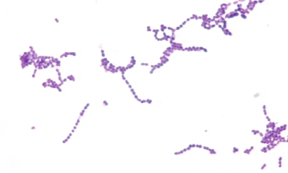
Preliminary labs were significant for metabolic acidosis on arterial blood gas: pH 7.00, pCO2 45, pO2 35 and HCO3 of 11. Upon presentation to the emergency room he was placed on BiPAP. Soon after BiPAP was begun he had a witnessed generalized tonic-clonic seizure, which responded to lorazepam. After stabilization, he was intubated and admitted to the ICU, and his acidosis was managed. Initial workup was focused on a possible PE given the significant tachypnea, tachycardia and respiratory failure. Preliminary chest x-ray showed no acute cardiopulmonary changes. A CT Angiography study of his chest did not reveal signs of possible PE. A head CT was ordered and revealed a ring enhancing 22 x 20 x 17 mm mass in the subcortical superior lateral right frontal lobe at the level of coronal suture. At this time, it seemed possible that the intracranial mass was caused by metastatic disease given the patients history of hepatitis C. Workup for malignancy consisted of a CT of his Chest, Abdomen and Pelvis, all of which were negative for signs of a primary malignancy. At this time, neurosurgery was consulted and an MRI with and without contrast was ordered, which revealed a 28 x 22 x 26 mm mass with surrounding edema in the anterior superior lateral right frontal lobe and a smaller 11 x 11 x 10 mm ring enhancing lesion with surrounding edema in the right head of the caudate. The MRI findings were most consistent with brain abscesses. Infectious Disease was consulted and the patient was started on empiric IV ceftriaxone and metronidazole. The frontal lobe abscess was then successfully incised and drained by neurosurgery utilizing stealth needle technique, and then irrigated with vancomycin. Cultures from the abscess drainage were positive for Viridans streptococci (Figure 1).
The patient's neurologic status and acidosis gradually improved and he was successfully extubated. Once the patient was able to provide history, he reported that he had undergone two deep cleaning dental procedures for severe periodontal disease approximately 6 months prior to this admission. The brain abscess was likely a result of oral seeding stemming from the dental procedures. This is especially likely given the culture results in the setting of ongoing prednisone and infliximab immunosuppression. He was discharged home on ceftriaxone and metronidazole. At his neurosurgery follow-up appointment, a repeat CT was ordered, and showed a rim-enhancing right frontal 17 x 14 mm lesion, located at the gray-white matter junction with expected post-surgical changes from his right frontal craniotomy. He had complete return of motor function without lasting neurological deficits or seizures. Immunosuppressive therapy for his Crohn's Disease was not restarted. After six months of follow-up, the patient is doing well without signs of neurologic deficits, systemic infection, or Crohn's Disease recurrence.
Discussion
In a patient with focal neurologic signs and multiple brain lesions on imaging, the two most important items on the differential diagnosis are infection and metastatic disease. The two conditions are commonly differentiated by findings in the history as well as specific imaging findings unique to each condition, though there can be considerable overlap in the features that make preoperative diagnosis difficult in some cases. A classic history in a patient with a brain abscess contains an obvious source of infection and presence of fever or chills, while a classic history in a patient with brain metastasis may include cachexia, and historical or current primary malignancy. Imaging of the brain via CT and MRI is known to show a rim-enhancing mass with perifocal edema in both pyogenic brain abscess, and cystic or necrotic metastasis, making a precise diagnosis challenging in select cases [3]. While the gold standard in differentiating a cerebral abscess from metastasis is considered to be an MRI with a diffusion sequence, an MRI scanner is not always available [4].
As our patient had a long history of uncontrolled Hepatitis C, the possibility of metastatic hepatocellular carcinoma was considered. In the United States, Hepatitis C is the most common cause of hepatocellular carcinoma [5]. Hepatitis C is known to lead to hepatocellular carcinoma by the mechanisms of chronic inflammation, cellular death and proliferation, and direct oncogenic potential [5]. However, hepatocellular carcinoma causing brain metastasis is relatively rare, with reported frequencies ranging from 0.2% to 2.2% at autopsy [6].
Cerebral abscesses are localized areas of suppuration that develop within the brain parenchyma [7]. They are known to occur following cranial trauma or surgery, direct extension from an existing infection, or via hematogenous seeding [8]. Location of cerebral lesions can be an important tool to discern the cause of the infection: otitis media and mastoiditis are associated with abscesses in the inferior temporal lobe and cerebellum, while infections of the frontal sinus, ethmoid sinus, and oral cavity are known to cause infectious lesions in the frontal lobe [7]. In our patient, the most likely mechanism is hematogenous seeding because he has multiple brain lesions, and due to the specific bacteria isolated from his abscesses [9].
The most common bacteria found in cerebral abscesses are Viridans streptococci, and Staphylococcus aureus [10]. Classically, Viridans streptococci are associated with dental infections, while S. aureus is associated with skin infections and intravenous drug use. Our patient's history of dental procedures and finding of Viridans streptococci align. However, in an immunocompromised host such as our patient, a multitude of organisms are possible, including fungi [11]. The medications causing immunosuppression in our patient are infliximab and prednisone, which are associated with cerebral infections caused by Toxoplasma gondii [12], Listeria monocytogenes [13], Nocardia spp.[14], and Aspergillus spp.[15], and many other pathogens. Following treatment for cerebral abscess for a patient on immunosuppression, there is often debate on the safety of restarting immunomodulating medications. In patients with severe autoimmune disease requiring immunomodulators, several authors have advocated for restarting the immune modulating medication in addition to a chronic suppressive antibiotic [16]. In our patient, however, immunomodulators were not restarted, and the patient has not developed signs of Crohn's disease recurrence.
Figure 2
1 month postoperative Head CT, showing a rim-enhancing right frontal 1.7 x 1.4 cm lesion, located at the gray-white matter junction with expected post-surgical changes following right frontal craniotomy. Preoperative images are not available.

Conclusions
In a patient with a history of immunosuppression and a recent dental procedure presenting with focal neurologic signs and fever, cerebral abscess should be at the top of the differential diagnosis.
Consent
Written informed consent was obtained from the patient for publication of this case report and any accompanying images. A copy of the written consent is available for review by the Editor-in-Chief of this journal.
Authors' Contributions
DC prepared the abstract, introduction, and discussion. RW collected the case information and assembled the case report.
References
- Mamelak AN, Mampalam TJ, Obana WG, Rosenblum ML. Improved management of multiple brain abscesses: a combined surgical and medical approach. Neurosurgery. 1995; 36: 76-85.
- Tan K, Patel S, Gandhi N, Chow F, Rumbaugh J, Nath A. Burden of neuroinfectious diseases on the neurology service in a tertiary care center. Neurology. 2008; 71: 1160-6. Doi: 10.1212/01.wnl.0000327526.71683.b7.
- Toh CH, Wei KC, Chang CN, Ng SH, Wong HF, Lin CP. Differentiation of brain abscesses from glioblastomas and metastatic brain tumors: comparisons of diagnostic performance of dynamic susceptibility contrast-enhanced perfusion MR imaging before and after mathematic contrast leakage correction. PLoS One. 2014; 9: e109172. Doi: 10.1371/journal.pone.0109172.
- Rees J. Advances in magnetic resonance imaging of brain tumours. Curr Opin Neurol. 2003; 16: 643-50.
- Luis Jesuino de Oliveria Andrade, Argemiro D'Oliveira, Junior, Rosangela Carvalho Melo,1 Emmanuel Conrado De Souza,1 Carolina Alves Costa Silva, and Raymundo Parana. Association Between Hepatitis C and Hepatocellular Carcinoma. J Glob Infect Dis. 2009; 1: 33-37. Doi: 10.4103/0974-777X.52979.
- Jiang XB, Ke C, Zhang GH, Zhang XH, Sai K, Chen ZP, et al. Brain metastases from hepatocellular carcinoma: clinical features and prognostic factors. BMC Cancer. 2012; 12: 49. Doi: 10.1186/1471-2407-12-49.
- Corson MA, Postlethwaite KP, Seymour RA. Are dental infections a cause of brain abscess? Case report and review of the literature. Oral Dis. 2001; 7: 61-5.
- Brouwer MC, Tunkel AR, McKhann GM, van de Beek D. Brain Abscess. N Engl J Med. 2014; 371: 447-56. Doi: 10.1056/NEJMra1301635.
- Bakshi R, Wright PD, Kinkel PR, Bates VE, Mechtler LL, Kamran S, et al. Cranial magnetic resonance imaging findings in bacterial endocarditis: the neuroimaging spectrum of septic brain embolization demonstrated in twelve patients. J Neuroimaging. 1999; 9: 78-84.
- Brouwer MC, Coutinho JM, van de Beek D. Clinical characteristics and outcome of brain abscess: systematic review and meta-analysis. Neurology. 2014; 82: 806-13. Doi: 10.1212/WNL.0000000000000172.
- Hagensee ME, Bauwens JE, Kjos B, Bowden RA. Brain abscess following marrow transplantation: experience at the Fred Hutchinson Cancer Research Center, 1984-1992. Clin Infect Dis. 1994; 19: 402-8.
- Young JD, McGwire BS. Infliximab and Reactivation of Cerebral Toxoplasmosis. N Engl J Med. 2005; 353: 1530-1; 1530-31.
- Chen FW, Matar W,Hersch M. Listeriosis Complicating Infliximab Treatment in Crohn's Disease. J ClinGastroenterolTreat 2016, 2: 024
- Valarezo J, Cohen JE, Valarezo L, Spektor S, Shoshan Y, Rosenthal G, et al. Nocardial cerebral abscess: report of three cases and review of the current neurosurgical management. Neurol Res. 2003; 25: 27-30.
- Erdogan E, Beyzadeoglu M, Arpaci F, Celasun B. Cerebellar aspergillosis: case report and literature review. Neurosurgery. 2002; 50: 874-6; 876-7.
- Abreu C, Rocha-Pereira N, Sarmento A, Magro F. Nocardia infections among immunomodulated inflammatory bowel disease patients: A review. World J Gastroenterol. 2015; 21: 6491-8. Doi: 10.3748/wjg.v21.i21.6491.
Authors submit all Proposals and manuscripts via Electronic Form!


