Case Report
Partial Transection and Healing of the Right Main Bronchus: an Unreported Complication of Chest Tube Insertion
Tarig E. Fadelelmoula1*, Momen M. Abdalla2 and Husameldin S. Hussein2
1The Department of Respiratory Care, University of Almaarefa, Riyadh, Saudi Arabia
2Department of Chest Medicine, Al Shaab Teaching Hospital, Khartoum, Sudan
*Address for Correspondence: Tarig E. Fadelelmoula, The Department of Respiratory Care, University of Almaarefa, Riyadh, Saudi Arabia, Tel: +966-542-521-796; E-mail: ttoom@mcst.edu.sa/eltarig70@hotmail.com
Dates: 07 July 2018; Approved: 21 August 2018; Published: 22 August 2018
Citation this article: Fadelelmoula TE, Abdalla MM, Hussein HS. Partial Transection and Healing of the Right Main Bronchus: an Unreported Complication of Chest Tube Insertion. Int J Case Rep Short Rev. 2018;4(2): 035-037.
Copyright: © 2018 Fadelelmoula TE, et al. This is an open access article distributed under the Creative Commons Attribution License, which permits unrestricted use, distribution, and reproduction in any medium, provided the original work is properly cited.
Keywords: Chest tube; Thoracotomy; Trauma; Bronchus; Trocar
Abstract
Chest tube is the most commonly performed surgical procedure in chest surgery. The lung is the most commonly injured organ during chest tube placement. Published complications in adult patients included lacerations of the lung, intercostal artery, esophagus, stomach, liver, spleen, diaphragm, pulmonary artery and atrium as well as right ventricular compression, but to our knowledge this is first case reported with Partial transection and healing of the main bronchus following chest tube insertion. We report a 35 years old male referred to our hospital with chest pain and worsening shortness of breath and history of chest pain and mild haemoptysis immediately following chest tube insertion. We conclude that lung damage during chest tube insertion is common, but can be avoided by using blunt dissection technique without the use of a trocar; trainee should be supervised while performing such a common procedure and that post tube thoracotomy care may foster early detection and halts advancement of those complications.
Introduction
Chest tube is the most commonly performed surgical procedure in chest surgery. As a vital surgery that saves lives, general surgeons, intensivists, emergency doctors, and pulmonologists, may at one time or the other, be required to perform chest tube insertion procedure. The first documented description of a closed chest tube drainage system for the drainage of empyema was by Hewet [1]. The lung is the most commonly injured organ during chest tube placement. Patients with underlying chest pathologies like decreased lung compliance, consolidation of the underlying parenchyma or marked pleural adhesions are at a higher risk for laceration. These conditions limit normal displacement of the lung when confronted by the chest tube. The use of a trocar and the inability to sufficiently visualize the pleural space prior to tube placement also increase the risk of chest tube related lung laceration [2-4]. Published complications in adult patients included lacerations of the lung, intercostal artery, esophagus, stomach, liver, spleen, diaphragm, pulmonary artery and atrium as well as right ventricular compression [5-9]. To our knowledge, there is no reported case where the main bronchus was partially transected during chest tube insertion. We report the first case of partial transection and healing of the right main bronchus as a complication of chest tube insertion.
Case Report
We report a 35 years old male with partial transection and healing of the right main bronchus following chest tube thoracotomy. He was referred to Alshaab Teaching Hospital with chest pain and worsening shortness of breath, pulse rate was135/min, respiratory rate was 33/min, SpO2 was 85% on room air and BP was 100/65 mmHg. He gave history of increasing dyspnoea, chest pain and mild haemoptysis immediately following chest tube insertion for massive right side traumatic pleural effusion four weeks prior to referral. On physical examination, the patient was distressed and his chest examination was consistent with right lung collapse, other systems were normal.
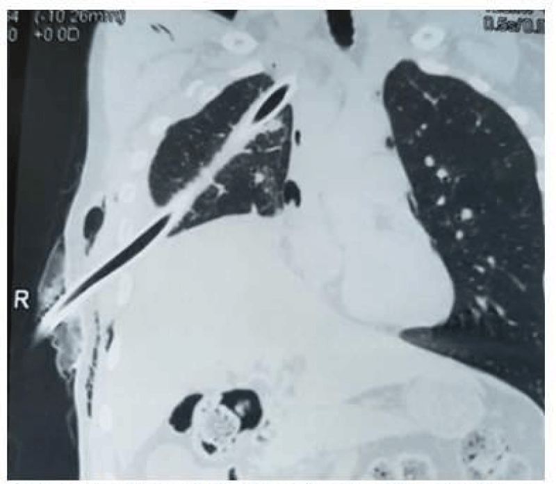
Imaging was done and x-Ray of his chest revealed just opacified right hemithorax with loss of volume (Figure 1). CT-Scan showed the chest tube penetrating the right lung up to just prior to trachea completely traversing the right main bronchus and there is right lung volume loss (Figure 2). Bronchoscopy revealed a healed stump of the right main bronchus confirming that partial transaction was followed with complete healing (Figure 3). Diagnosis of partial transection and healing of the right main bronchus suspected and the patient was advised to travel abroad where a complex thoracic surgery for bronchus reconstruction and lung expansion was successfully performed.
Discussion
Thoracic trauma is commonly treated with chest tube insertion and it is lifesaving in severely injured patients. The overall complication rate associated with this procedure is up to 30% among all operators [10]. For the prevention of iatrogenic injuries, it is recommended that all chest tubes should be inserted in the “triangle of safety”. Clinical guidelines also suggest that the placement of a chest drain outside the “triangle of safety” should always be performed or discussed with a senior expert clinician. Guidelines also emphasise on proper training of technique [11]. Following chest tube insertion, good nursing care is needed because the chest tube can migrate. A postoperative chest radiograph is required to confirm the chest-tube position. If the chest tube is in the correct position, then the water column in the water-seal chamber moves during respirations. The column will not move when the lung is fully expanded. In the case we are reporting, the wrong position of the tube was immediately evident from the early post tube insertion symptoms, namely chest pain, dyspnoea and haemoptysis and emergency surgical care should have been instituted to manage immediate complications and prevent late complications of the procedure. Surgical care usually aims at hemostasis and thoracotomy for early repair of injuries caused by the tube. Although check chest X-ray was done but referral to tertiary hospital was delayed.
Conclusion
Lung damage during chest tube insertion is common but to our knowledge this is first case reported with partial transection and healing of the main bronchus following chest tube insertion. To prevent these complications, blunt dissection technique without the use of a trocar should be used; trainee should be supervised while performing such a common procedure and that post tube thoracotomy care may foster early detection and halts advancement of those complications.
References
- Hewett FC. Thoracentesis: The Plan of continuous aspiration. Br Med J. 1876; 1: 317. https://goo.gl/SQqYQj
- Miller KS, Sahn SA. Chest tubes. Indications, technique, management and complications. Chest. 1987; 91: 258-264. https://goo.gl/QLgV8z
- Fraser RS. Lung perforation complicating tube thoracostomy: Pathologic description of three cases. Hum Pathol. 1988; 19: 518-523. https://goo.gl/q4YszH
- Meisel S, Ram Z, Priel I, Nass D, Lieberman P. Another complication of thoracostomy-perforation of the right atrium. Chest. 1990; 98: 772-773. https://goo.gl/HDf7kp
- Kollef MH, Dothager DW. Reversible cardiogenic shock due to chest tube compression of the right ventricle. Chest. 1991; 99: 976-980. https://goo.gl/hUDFbo
- McFadden PM, Jones JW. Tube thoracostomy: anatomical considerations, overview of complications, and a proposed technique to avoid complications. Mil Med. 1985; 150: 681-685. https://goo.gl/ueWkQf
- Shapira OM, Aldea GS, Kupferschmid J, Shemin RJ. Delayed perforation of the esophagus by a closed thoracostomy tube. Chest. 1993; 104: 1897-1898. https://goo.gl/UQHjVi
- Singh KJ, Newman MA. Pulmonary artery catheterization: an unusual complication of chest tube insertion. Aust N Z J Surg. 1994; 64: 513-514. https://goo.gl/GzCPJb
- Ball CG, Lord J, Laupland KB, Gmora S, Mulloy RH, Ng AK, et al. Chest tube complications: how well are we training our residents. Can J Surg. 2007; 50: 450-458. https://goo.gl/eo6hDM
- Elsayed H, Roberts R, Emadi M, Whittle I, Shackcloth M. Chest drain insertion is not a harmless procedure--are we doing it safely. Interact Cardiovasc Thorac Surg. 2010; 11: 745-748. https://goo.gl/rjhE2Z
- Gossage JA, Chukwuemeka AO, Dussek JE. Intercostal drain migration post esophagectomy. Dis Esophagus. 2003; 16: 268-269. https://goo.gl/JQfT8A
Authors submit all Proposals and manuscripts via Electronic Form!


