Maheshgir S. Gosavi*
Siraj Hospital, Senior Consultant, Thane, India
*Address for Correspondence: Maheshgir S. Gosavi, Siraj Hospital, Vanjarpatti Naka, Bhiwandi, Thane, India, Tel: +910-252-225-3200/+918-605-870-884; E-mail: raddon84@rediffmail.com/raddon84@gmail.com
Dates: 05 April 2018; Approved: 21 June 2018; Published: 26 June 2018
Citation this article: Gosavi MS. Unusual Tubal Twin Ectopic Gestation - Report of Three Different Cases. Int J Case Rep Short Rev. 2018;4(2): 031-034.
Copyright: © 2018 Gosavi MS. This is an open access article distributed under the Creative Commons Attribution License, which permits unrestricted use, distribution, and reproduction in any medium, provided the original work is properly cited.
Keywords: Spontaneous; Tubal twin ectopic; Transvaginal usg
Abstract
Gestation outside the uterine cavity in which the implantation occurs in any tissue other than the endometrium is referred as ectopic pregnancy. The most place for occurring ectopic pregnancy (97% of cases) is the fallopian tubes including ampulla (55%), isthmus (25%), and fimbria (17%), and in 3% of patients ectopic pregnancy occurs in the abdominal cavity, ovary, or cervix. The tubal twin ectopic pregnancy is a rare condition, and the first unilateral tubal twin was reported by De Ott in 1891, and the first live twin tubal ectopic pregnancy was reported in 1944. A live tubal twin ectopic pregnancy is a very rare condition and among >100 reports of tubal twin pregnancies, till now, only 8 cases were live. Early diagnosis and treatment of women with tubal twin ectopic pregnancy is very important and may decrease the risk of tubal rupture. I present three cases of tubal twin ectopic gestation. In the first case, spontaneous unilateral live tubal twin ectopic gestation. The second and third cases spontaneous ruptured twin ectopic gestation. All three cases were successfully managed and there was no history of assisted reproductive technique fertilization or pelvic inflammatory disease.
Introduction
Gestation outside the uterine cavity in which the implantation occurs in any tissue other than the endometrium is referred as ectopic pregnancy. The most place for occurring ectopic pregnancy (97% of cases) is the fallopian tubes including ampulla (55%), isthmus (25%), and fimbria (17%), and in 3% of patients ectopic pregnancy occurs in the abdominal cavity, ovary, or cervix [1]. The tubal twin ectopic pregnancy is a rare condition, and the first unilateral tubal twin was reported by De Ott in 1891, and the first live twin tubal ectopic pregnancy was reported in 1944 [2]. A live tubal twin ectopic pregnancy is a very rare condition and among >100 reports of tubal twin pregnancies, till now, only 8 cases were live [3]. Early diagnosis and treatment of women with tubal twin ectopic pregnancy is very important and may decrease the risk of tubal rupture. The present report describes a successful management of spontaneous twin tubal ectopic pregnancies with no history of assisted reproductive technique fertilization or pelvic inflammatory disease.
Case 1
Case presentation
A 35 year old female, came to casualty at midnight with pain in abdomen since three days with 8 weeks of amenorrhea and bleeding per vaginum since morning .She had one living issues with full term normal deliveries. She had history of two spontaneous abortions. Upon examination, her vitals were stable. Abdomen examination was unremarkable. Upon per vaginum examination the cervical movement was tender, the uterus was bulky and soft, and bogginess and tenderness were felt in the right adnexa. However, her urine pregnancy test was positive.
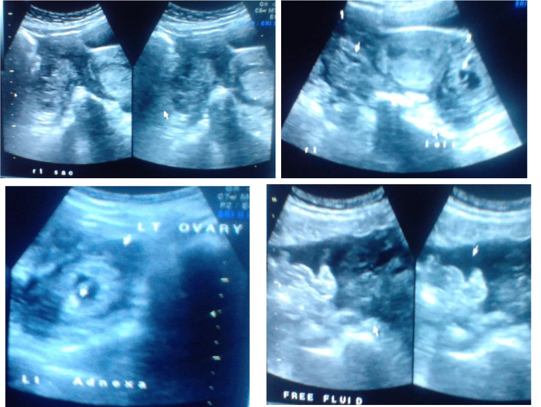
Ultrasonography with doppler study showed a uterus of 8.3 x 5.4 x 4.2 cms with endometrial thickness of 10 mm and there was evidence of large heterogeneous lesion in the right adnexa of a size 7.1 x 5.1 cms, with sac like structure within which two foetal poles measuring 1.4 cm = 7 weeks and five days. Fetal cardiac activity was appreciated. Mild free fluid was seen in the abdomen. An impression of live twin ectopic pregnancy in the right adnexa was made with mild free fluid in an abdomen. A pseudosac like structure was noticed in endometrial cavity. Both ovaries were visualised separately from the right adnexal mass.
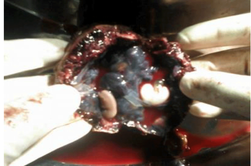
In view of the Twin live ectopic pregnancy she was immediately operated. Intraoperatively, the ampullary part of the right fallopian tube was found showed a sac like structure. A right salphingotomy was done. She was discharged on the 7th day. On cut section of the sac there was evidence of two embryos Pathological evaluation of the surgical specimen showed a diamniotic monochorionic twins. Pregnancy within the fallopian tube and measurement of the twin feti estimated their gestational age at 7 weeks.
Case 2
A 30 year old female, G2L1A0 came to casualty with severe pain in abdomen since two days with 6 weeks of amenorrhea and bleeding per vaginum since morning .She had one living issues with full term normal deliveries. Upon examination, her vitals were stable. Abdomen examination was unremarkable. However, her urine pregnancy test was positive.
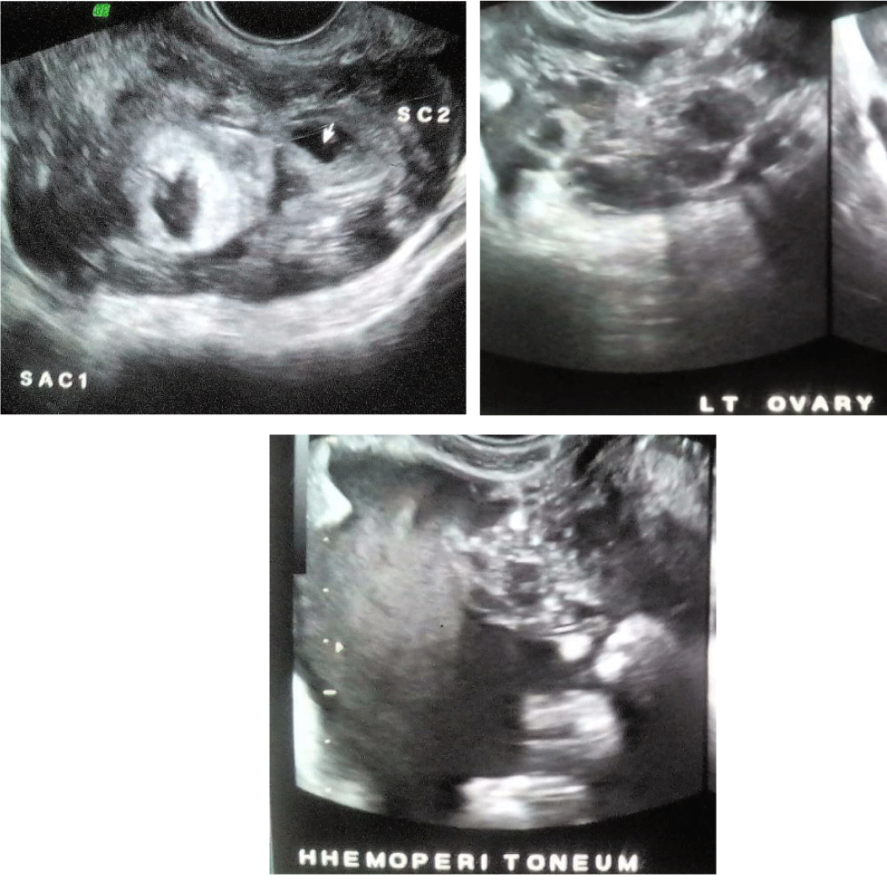
Spontaneous pregnancy there was no history of induction of ovulation by drugs or artificial reproductive techniques. The patient was taken for ultra sonography. Transvaginal Sonography showed a heterogeneous mass in right adnexa measuring approximately 7.3 x 4.6 cm The mass showed irregular Gestational sac measuring approximately 0.3 cm corresponding to 5 weeks and another irregular sac measuring 0.5 cm corresponding to 5weeks 2 days Right ovary was visualised separately from this mass.
Left ovary was visualised and appears normal .Moderate hemoperitoneum was seen .Uterus was bulky however there was no intrauterine gestational sac. The diagnosis of right adnexal ruptured ectopic gestation was made. Preliminary investigations were done and patient was taken up for laparotomy in view of right adnexal mass and hemoperitoneum. Per operatively gross morphology was: Right adnexal complex mass with moderate hemoperitoneum. Right salpingectomy was performed. Patient’s post-operative period was uneventful. Histopathology report came out to be right adnexal two gestational sacs were noted.
Case 3
A 36 year old female, G3L2A0 came to casualty with severe pain in abdomen since three to four days with 6 weeks of amenorrhea and bleeding per vaginum since morning. Upon examination, her vitals were stable. Abdomen examination was unremarkable. Upon per vaginum examination the cervical movement was tender, the uterus was bulky and soft, and bogginess and tenderness were felt in the both adnexae. However, her urine pregnancy test was positive. Spontaneous pregnancy there was no history of induction of ovulation by drugs or artificial reproductive techniques the patient was taken for ultra sonography. Transvaginal Sonography showed a heterogeneous mass in right adnexa with irregular Gestational sac measuring approximately 1.0 cm corresponding to 5 weeks 5 days Right ovary was visualised separately from this mass.
Left adnexa irregular Gestational sac measuring approximately 0.8cm corresponding to 5 weeks 4days. Left ovary was visualised separately from this mass. Mild to Moderate hemoperitoneum was seen. Uterus was bulky however there was no intrauterine gestational sac. The diagnosis of bilateral adnexal ruptured ectopic gestation was made. Preliminary investigations were done and patient was taken up for laparotomy in view of bilateral adnexal masses and hemoperitoneum. Per operatively gross morphology was: Bilateral adnexal complex masses with moderate hemoperitoneum. Bilateral salpingotomy was performed. Patient’s post-operative period was uneventful.
Discussion
The preoperative diagnosis of twin tubal pregnancy is difficult and potentially may lead to significant morbidity or mortality [4]. Based on the changing pattern of clinical presentation, the gynecologist should pay special attention in such suspected cases. All available methods of diagnosing the suspected cases should be used including ultrasonography and blood chemistry tests such as beta Human Chorionic Gonadotropin (hCG) level. In the present case, transabdominal sonography was performed and the initial diagnosis was unruptured ectopic pregnancy. The serum hCG level was not tested. Therefore, the twin tubal pregnancy was not in mind, even if it had a higher incidence of rupture. Her treatment was delayed and ruptured of tubal pregnancy occurred subsequently. There is evidence that the hCG level of twin tubal pregnancies is higher than that of a singleton tubal pregnancy and surgical treatment of those cases is appropriate [5]. The mean hCG level of twin and singleton ectopic pregnancies was 9,849 mIU/ml and less than 3,000 mIU/ml, respectively [3]. Transvaginal sonography has shown to be effective in the diagnosis of intact twin tubal pregnancy and extremely sensitive in the detection of free pelvic fluid [6]. The pathogenesis of unilateral twin tubal pregnancy is not clear. Several factors are thought to contribute to the occurrence of ectopic pregnancy. These include mechanical obstruction within the tube [7], defects of the zygote itself or in the hormonal milieu. It is likely that the twin tubal ectopic pregnancy is also increased in patients treated by in vitro fertilization [8,9], but the conception in the present case was spontaneous. The unilateral twin tubal pregnancy cans occur spontaneously [10]. Some investigators suggested that the larger cell mass of the fertilized twin zygote might cause retarded tubal transport along a damaged tube and result in tubal implantation [11]. The zygosity of twin in the present case was not studied. Most of the twin tubal pregnancies were thought to be monozygotic. However, in the majority of the reported cases, the zygosity was determined subjectively by using observation such as size and gestational age similarity, fetal membranes and number of corpus luteum [12]. It seems to be that the method to determine zygosity of the twins by using DNA probes that detect restriction fragment length polymorphisms more accurately [13]. Some researchers speculate that many of the unilateral ectopic twins who were thought to be monozygotic may actually have been dizygotic [14].
In the past, there have been a few cases of twin tubal pregnancies diagnosed preoperatively [3,14]. Almost all the reported cases were diagnosed retrospectively either by intra operation or histology [13]. Over the years, the treatment of ectopic pregnancy has progressed from salpingectomy by laparotomy to conservative surgery by laparoscopy and more recently, by medical therapy. There are a few reports of successful laparoscopic management of ruptured tubal twin pregnancy [15] and operative laparoscopic salpingostomy [16]. The laparotomy is better for this situation to control the bleeding point from the ruptured site. The present cases underscores the need of early recognition and accurate diagnosis of twin tubal pregnancy, that it may grow larger and therefore, have a higher risk of rupture. The high-resolution transvaginal sonography is very helpful in the diagnosis of this condition [15,17].
 Figure 1: Shows Right adnexal sac with Twin live ectopic gestation of approximately 7wks 5 days .No intrauterine pregnancy. Both ovaries are separately visualised.
 Figure 2: Pathological evaluation of the surgical specimen showed a diamniotic monochorionic twins pregnancy.
Conclusion
A spontaneous twin tubal pregnancy can occur in patients who have no known predisposing factor. Early diagnosis has made this disorder amenable to appropriate treatment. The high-resolution transvaginal sonography is very helpful in the diagnosis of this condition. Twin-ectopic gestations are extremely rare but there are treatment options. These have typically been classified as either conservative or surgical.
References
- Lozeau AM, Potter B. Diagnosis and management of ectopic pregnancy. Am Fam Physician. 2005; 72: 1707-1714. https://goo.gl/ji5Pfw
- Dede M, Gezginç K, Yenen M, Ulubay M, Kozan S, Guran S, et al. Unilateral tubal ectopic twin pregnancy. Taiwan J Obstet Gynecol. 2008; 47: 226-228.
- Eddib A, Olawaiye A, Withiam-leitch M, Rodgers B, Yeh J. Live twin tubal ectopic pregnancy. Int J Gynaecol Obstet. 2006; 93: 154-155. https://goo.gl/iPqoPm
- Sherer DM, Liberto L, Woods JR Jr. Preoperative sonographic diagnosis of a unilateral tubal twin gestation with documented fetal heart activity. J Ultrasound Med. 1990; 9: 729-731. https://goo.gl/BcLEzp
- Zakut H, Fiengold M, Lipnitzky W. Unilateral tubal pregnancy of twins. Clin Exp Obstet Gynecol. 1983; 10: 157-158. https://goo.gl/Pf3kqv
- Gabrielli S, Marconi R, Ceccarini M, Valeri B, De Iaco P, Pilu G, et al. Transvaginal and three dimensional ultrasound diagnosis of twin tubal pregnancy. Prenat Diagn. 2006; 26: 91-93.
- Ansari SM, Nessa L, Saha SK. Twin ectopic pregnancy 10 years after permanent sterilization. Australas Radiol. 2000; 44: 107-108. https://goo.gl/F9ALpv
- Dor J, Rudak E, Mashiach S, Goldman B, Nebel L. Unilateral tubal twin pregnancy following in vitro fertilization and embryo transfer. Fertil Steril. 1984; 42: 297-299. https://goo.gl/vAG7zL
- Goker EN, Tavmergen E, Ozcakir HT, Levi R, Adakan S. Unilateral ectopic twin pregnancy following an IVF cycle. J Obstet Gynaecol Res. 2001; 27: 213-215.https://goo.gl/QfiuPM
- Sur SD, Reddy K. Spontaneous unilateral tubal twin pregnancy. J R Soc Med. 2005; 98: 276. https://goo.gl/5geRJn
- Ash KM, Lyons EA, Levi CS, Lindsay DJ. Endovaginal sonographic diagnosis of ectopic twin gestation. J Ultrasound Med. 1991; 10: 497-500. https://goo.gl/rZVVsR
- Basama FM. Preoperative diagnosis of unilateral tubal twin ectopic pregnancy with one live twin. J Obstet Gynaecol. 2003; 23: 313-314. https://goo.gl/wF3E3U
- Neuman WL, Ponto K, Farber RA, Shangold GA. DNA analysis of unilateral twin ectopic gestation. Obstet Gynecol. 1990; 75: 479-483. https://goo.gl/JokHZd
- Parker J, Hewson AD, Calder-Mason T, Lai J. Transvaginal ultrasound diagnosis of a live twin tubal ectopic pregnancy. Australas Radiol. 1999; 43: 95-97. https://goo.gl/DKNFrs
- Gassibe EF, Gassibe E. Laparoscopic management of ruptured tubal twin pregnancy. J Am Assoc Gynecol Laparosc. 1996; 3: 15. https://goo.gl/gaQacC
- Shwayder JM, Mahoney V, Bersinger DE. Unilateral twin ectopic pregnancy managed by operative laparoscopy. A case report. J Reprod Med. 1993; 38: 314-316. https://goo.gl/J1QpxJ
- Gerscovich EO, McGahan JP. High-resolution ultrasonography in the diagnosis of twin tubal ectopic pregnancies. J Clin Ultrasound. 1991; 19: 501-504. https://goo.gl/MH46FD
Authors submit all Proposals and manuscripts via Electronic Form!


