Maheshgir S Gosavi*
Siraj Hospital, Senior Consultant, Thane, India
*Address for Correspondence: Maheshgir S.Gosavi, Siraj Hospital, Vanjarpatti Naka, Bhiwandi, Thane, India, Tel: +910-252-225-3200/+918-605-870-884; E-mail: [email protected]/[email protected]
Dates: 22 January 2018; Approved: 01 February 2018; Published: 03 February 2018
Citation this article: Gosavi MS. Myxoglobulosis of the Appendix: A Rare Cause of Pelvic Pain. Int J Case Rep Short Rev. 2018;4(1): 009-011.
Copyright: © 2018 Gosavi MS. This is an open access article distributed under the Creative Commons Attribution License, which permits unrestricted use, distribution, and reproduction in any medium, provided the original work is properly cited.
Keywords: Myxoglobulosis; Pelvic pain; Mucocele
Abstract
Myxoglobulosis, also known as caviar appendix, is an unusual and rare variant of appendix mucocele. I report a case of a 29-year-old woman who presented with a 6 day history of pelvic pain after pelvic ultrasound revealed an enlarged appendix. Mucocele alone does not constitute a rare finding in routine appendectomy. However, the coexistence of multiple small intraluminal globules, described in the past as “frog eggs” or “fish eggs”, constitutes a special type of mucocele described as “myxoglobulosis”.
Introduction
Myxoglobulosis, also known as caviar appendix, is an unusual and rare variant of appendix mucocoele. The initial description of myxoglobulosis of the appendix dates back to 1897 by Latham [1-3], nearly 55 years after the first description of appendiceal mucocele by Rokitanski [4]. I report a case of a 29-year-old woman who presented with a 6 day history of pelvic pain after pelvic ultrasound revealed an enlarged appendix. Macroscopically, the appendix had a proximal occlusive membrane and the lumen was distended by pearl-like cream white spheroids measuring 2–7 mm in diameter. Sectioning of the appendix revealed the presence in the dilated appendiceal lumen of numerous whitish opaque globules ranging in size from 0.2 to 0.7 cm in diameter. On microscopic examination, the globules consisted of faintly eosinophilic laminations of mucin surrounding an amorphous granular core. The mucin was identified by positivity with histochemical mucin stains. After thorough microscopic examination of the appendix, our case was diagnosed as myxoglobulosis .The features of myxoglobulosis will be discussed as well as a brief review of the relevant literature.
Case Presentation
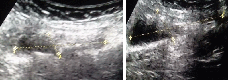
I report a case of a 29-year-old woman who presented with a 6 day history of pelvic pain and amenorrhea for 6 weeks .On examination there was tenderness with palpable lump in right iliac fossa. On Ultrasound examination tubular, non-compressible cystic structure in seen in right iliac fossa and extending into pelvis on right side .It shows multiple tiny echogenic foci within. Surrounding mesentery was inflamed .Uterus and both ovaries were normal. Both adnexa was clear .No evidence of intrauterine or extra uterine gestational sac.
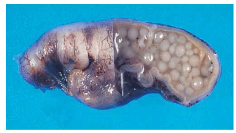
The differential diagnosis includes intraperitoneal or extra peritoneal lesions. Intraperitoneal masses to consider include ovarian cysts and tumours, duplication cysts, mesenteric and omental cysts, mesenteric hematoma or tumour, and abdominal abscess. Of the retroperitoneal disorders to be considered, retroperitoneal inflammation, tumour or haemorrhages are important. Urine pregnancy test was done found to be negative. Routine preoperative test was done Patient was diagnosed of appendicitis with mucocoele and taken for laparoscopic appendectomy The pathology specimen consisted of a dilated appendix measuring 6 cm in length and 2 cm in diameter, adherent to the mesoappendix. On section of the appendix, multiple small globular egg-like structures escaped from both the cut ends. They were found to be colaced with some kind of mucinous jelly-like substance. Sectioning of the appendix revealed the presence in the dilated lumen of numerous whitish opaque globules ranging in size from 0.2 to 0.7 cm in diameter. The mesoappendix near the distal portion of the appendix showed mucin deposits and globules.
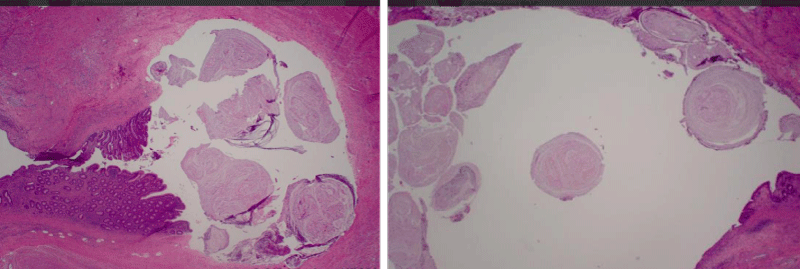
The entire appendix was submitted for histological evaluation. Hematoxylin-Eosin and histochemical stains Mucicarmine, Alcian Blue pH 2.5, PAS, and Diastase-PAS were performed. Histologically, the globules consisted of faint eosinophilic laminations of mucin surrounding an amorphous granular core. All of the globules stained uniformly positive with mucicarmine, Alcian Blue and PAS and were diastase resistant Focal gland serration and increased mucin-producing cells were found in the upper half of the mucosa, instead of slender filiform villi that are usually observed. In serial sections examined microscopically, the periappendiceal mucin did not contain any epithelial elements.
Discussion
Myxoglobulosis is an unusual variant of appendiceal mucocele [1-3]. The initial description of myxoglobulosis of the appendix dates back to 1897 by Latham [1,2], nearly 55 years after the first description of appendiceal mucocele by Rokitanski [4]. Since then at least 68 cases have been reported [1]. The incidence of appendiceal mucocele is estimated as 0.2-0.3 % of appendicectomy specimens, with myxoglobulosis constituting 0.35-0.8 % of mucoceles [1,5,6]. The factors leading to the transformation of mucin into globular bodies of myxoglobulosis are unknown. Various hypotheses have focused on the formation of a core, which then acts as a nidus for the concentric deposition of mucin, as the initiating event in the pathogenesis of the globules. Some authors propose the hypothesis of bacterial and necrotic epithelial debris origin and small mucinous masses putatively formed in dilated glandular crypts [1,7]. Lubin and Berle [2] in a report of two cases proposed that the core represented an organizing mass of mucin and granulation tissue originating in the appendiceal wall that broke off and underwent necrosis. In our case, examination of all the globules revealed inflammatory cellular elements. The same general mechanisms requisite in the production of appendiceal mucocele are involved in the pathogenesis of myxoglobulosis [8-10]. These mechanisms include (a) partial or complete obstruction of the lumen of the appendix and (b) continued mucin production by a normal or altered-for example, neoplastic-epithelium [11].
Most cases of myxoglobulosis have been incidental findings at autopsy or laparotomy. Patients with myxoglobulosis present clinically in a similar manner to those with mucocele of the appendix, the disorder occurring most commonly in the sixth or seventh decades of life with a female preponderance [4]. Patients are asymptomatic and myxoglobulosis is found incidentally at necropsy or laparotomy for other reasons [1,2,12]. In a few cases, patients present with episodic right lower abdominal pain with or without features of acute appendicitis as in our case [1,3,12]. Complications of myxoglobulosis are the same as mucocele and include intussusception, bleeding, perforation, peritonitis and pseudomyxoma peritonei [1,4,5,12-14]. Aroukatos et al. [15] reported that a case of myxoglobulosis of the appendix is associated with a ruptured diverticulum. Falah et al. [16] have reported a case of appendiceal myxoglobulosis associated with peritonitis due to perforated peptic ulcer. Padhy et al. [17] have reported a case of myxoglobulosis of the appendix. Routine histopathological examination is essential to diagnose myxoglobulosis.
Plain radiographs may show annular, non-laminated calcification of the globules [2,12], and a barium enema may show a smooth polypoid filling defect in the cecum, with features of an intramural extra mucosal lesion [1,4,6, 12]. Colonoscopy may show a smooth glassy submucosal or extra mucosal caecal mass moving in and out with respiratory movement; this endoscopic sign has been described as the trapped balloon sign [4]. Ultrasound and computerized tomography can be complementary in establishing the diagnosis [6]. The clinical and radiological differential diagnoses include other lesions of the appendix, such as polypoid adenoma, adenocarcinoma, lipoma, lymphoma, prolapse and intussusceptions [13,15]. Treatment is essentially by appendicectomy, but in cases of rupture or suspected malignancy a standard right hemicolectomy is needed [2-4,9,18].
 Figure 1: Ultrasound image showing cystic structure in right iliac fossa suggestive of appendix with multiple echogenic foci.
 Figure 2: Gross specimen of appendix showing multiple opaque globules. The appendiceal lumen is filled with pearl- or fish egg-like mucin globules.
 Figure 3: On histopathological examination, the globules consisted of faintly eosinophilic laminations of mucin surrounding an amorphous granular core were found.
Conclusion
Myxoglobulosis is an unusual variant of appendiceal mucocele .Diagnosis of appendicitis should be always be kept even in cases of pelvic pain .In our case with patients clinical history diagnosis of ectopic pregnancy was first suspected.
References
- Gonzalez JEG, Hann SE, Trujillo YP. Myxoglobulosis of the appendix. Am J Surg Pathol. 1988; 12: 962-966. https://goo.gl/52eYaU
- Lubin I, Berle E. Myxoglobulosis of the appendix. Report of two cases. Arch Pathol. 1972; 94: 533-536. https://goo.gl/ZxTRGt Rolon PA. Myxoglobulosis of the appendix. Int Surg. 1977; 62: 355-356. https://goo.gl/Jx3MyZ
- Rajman I, Leong S, Hassaram S, Marcon NE. Appendiceal mucocele: endoscopic appearance. Endoscopy. 1994; 26: 326-328. https://goo.gl/dVRhPw
- Madwed D, Mindelzun R, Jeffrey RB, Jr. Mucocele of the appendix: 11 imaging findings. Am J Radio. 1992; 159: 69-72. https://goo.gl/jcB8uL
- Issacs KL, Warshaner DM. Mucocele of the appendix: computed tomographic, endoscopic and pathologic correlation. Am J Gastroenterol. 1992; 87: 787-789. https://goo.gl/roZaj3
- Probstein JG, Lassar GN. Mucocele of the appendix, with myxoglobulosis. Ann Surg. 1948; 127: 171-176. https://goo.gl/y6hyPH
- Collins DC. A study of 50,000 specimens of the human vermiform appendix. Surg Gynecol Obstet. 1955; 101: 437-445. https://goo.gl/nuQT4N
- Pai RK, Beck AH, Norton JA, Longacre TA. Appendiceal mucinous neoplasms: clinic pathologic study of 116 cases with analysis of factors predicting recurrence. Am J Surg Pathol. 2009; 33: 1425-1439. https://goo.gl/9KrSJC
- Hollstrom E. Myxoglobulosis appendicis. Acta Chir Scand. 1941; 35: 347-359.
- Viswanath YK, Griffiths CD, Shipsey D, Oriolowo A, Johnson SJ. Myxoglobulosis, a rare variant of appendiceal mucocele, occurring secondary to an occlusive membrane. J R Coll Surg Edinb. 1998; 43: 204-206. https://goo.gl/2mwy5N
- Huang HC, Lui IP, Jeng KS. Intussusception of mucocele of appendix: a case report. Chin Med J. 1994; 53: 120-123. https://goo.gl/pzHHL5
- Brustmann H. Myxoglobulosis of the appendix associated with a proximal carcinoid and a pseudodiverticulum. Ann Diagn Pathol. 2006; 10: 166-168. https://goo.gl/nzhDLL
- Guionmet N, Martí E, Bella MR, Moreno A. Myxoglobulosis of the cecal appendix. Rev Esp Enferm Dig. 1990; 77: 365-367. https://goo.gl/h3fdDD
- Aroukatos P, Verras D, Vandoros GP, Repanti M. Myxoglobulosis of the appendix: a case associated with ruptured diverticulum. Case Rep Med. 2010; 2010. https://goo.gl/SaQN4y
- Falah HH, Maafi AA, Chatrnour G, Jahromi SK, Ebrahimi H. Myxoglobulosis, a rare variant of appendiceal mucocele: case report and review of the literature. Thrita J Med Sci. 2013; 2: 155-158. https://goo.gl/UCtJDP
- Padhy BP, Panda SK. Myxoglobulosis of Appendix A Rare Entity. Indian J Surg. 2013; 75: 337-339. https://goo.gl/efspmj
- Koçak C, Zengin AI, Kartufan FF, Metineren MH. Myxoglobulosis in the appendix. Turk J Surg. 2017; 33: 308-310. https://goo.gl/CRBXNX
Authors submit all Proposals and manuscripts via Electronic Form!


