Case Report
Temporary Pacemaker Induced Ventricular Fibrillations in Cath Lab Due to R on T Phenomenon
Sachin Sondhi1*, Rajesh Sharma1, Rajeev Merwaha1, Kunal Mahajan1, Ayushi Mehta2 and Munish Dev1
1Department of Cardiology, IGMC Shimla, HP, India
2Department of Anaesthesia, IGMC Shimla, HP, India
*Address for Correspondence: Sachin Sondhi, DM Cardiology Resident, Department of Cardiology, IGMC, Shimla, HP 171001, India, Tel: +918-219-508-161; E-mail: [email protected]
Dates: 13 December 2017; Approved: 26 January 2018; Published: 29 January 2018
Citation this article: Sachin S, Rajesh S, Rajeev M, Kunal M, Mehta A, et al. Temporary Pacemaker Induced Ventricular Fibrillations in Cath Lab Due to R on T Phenomenon. Int J Case Rep Short Rev. 2018;4(1): 006-008.
Copyright: © 2018 Sachin S, et al. This is an open access article distributed under the Creative Commons Attribution License, which permits unrestricted use, distribution, and reproduction in any medium, provided the original work is properly cited.
Keywords: Temporary Pacemaker Complications; R on T Phenomenon
Abstract
Temporary pacing is life saving and is established treatment of bradyarrhythmias developed after myocardial infarction. However, under sensing of QRS complex from ischemic area of ventricular cavity leads to inappropriate pacing spike on ST segment or on T wave of intrinsic complex, fall of pacing spike on this vulnerable period leads to induction of life threatening ventricular fibrillations. This phenomenon is known as “R on T phenomenon”. We report a case of 56 year male, presented to us with ST segment elevated inferior wall myocardial infarction with right ventricle involvement. After coronary angiography and prior to angioplasty, temporary pacemaker inserted and just after pacemaker insertion, he suddenly developed ventricular fibrillations. After getting reverted to sinus rhythm for few seconds with DC shock, these VF episodes were reoccurring at frequent intervals. On analyzing electrocardiogram on monitor, it was thought that temporary pacemaker was the culprit for this life threatening arrhythmia because of R on T phenomenon and pacemaker was turned off. After that he didn’t get any further episode of VF and successful PTCA of right coronary artery was performed.
Case Report
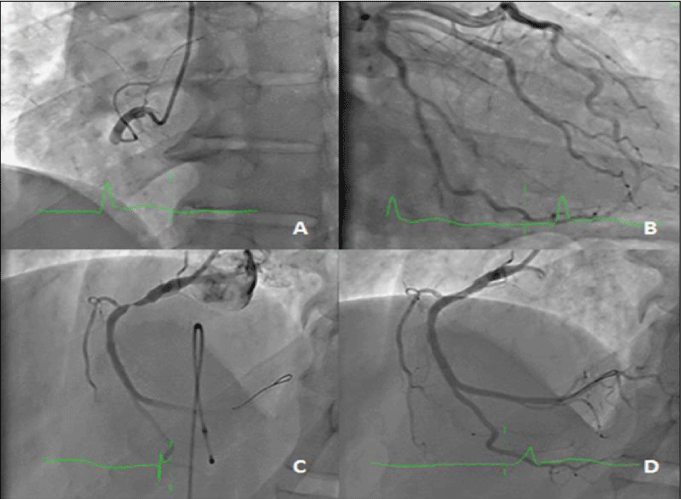
56 year male with no previous co morbidity presented to us within 2 hours of ST elevated inferior wall myocardial infarction with right ventricle involvement. At presentation, his BP was 90/60 mmHg, HR was 58/min, JVP was raised and chest was clear. He was immediately shifted to cath lab for primary PCI. His coronary angiography revealed total cut-off of RCA with normal left coronary circulation (Figure 2A,2B). In view of having sinus bradycardia temporary pacemaker was inserted and pacing lead was positioned in right ventricle apex.
Case 1
Case Presentation
As this patient requires emergent angioplasty, we only checked the pacing threshold and not the sensitivity threshold. The pacing threshold was 1.3mV, output was 5mV and sensitivity was 3mV. Immediately after pacing lead insertion patient became unstable and there were multiple episode of ventricular fibrillations. Every episode was reverted to sinus rhythm for few seconds with DC shock but it reoccurred again. Then bolus dose of amiodarone was given followed by multiple DC shocks every time, still rhythm was not reverted. It was noted on the telemetry that pacing spike falls on the T wave of normal intrinsic QRS complex which leads these repeated episodes of VF (Figure 1). The diagnosis of R on T phenomenon was made and pacemaker was turned off. After that no further episode of VF reoccurred. Then successful PCI was done and drug eluting stent was placed in RCA (Figure 2C,2D).
 Figure 1: R on T phenomenon demonstrated on recorded electrocardiogram. Note ST segment elevation in lead 2, after insertion of temporary pacing lead, there is inadequate sensing of intrinsic QRS complex leading to the pacemaker spike (vertical downward arrow) to fall on T wave and followed by paced complex, next complex is normal intrinsic complex and under sensing of this complex leads to pacing spike (vertical upward arrow) to fall on ST segment and development of ventricular fibrillations.
 Figure 2: (A) Showing RCA total cut off after the origin (B) Left coronary artery is normal (C) After crossing wire there good ante grade flow in RCA with 99% stenosis in proximal part, note pacing lead in right ventricle (D) Final result after DES placement in proximal RCA with no residual stenosis.
Discussion
The “R-on-T phenomenon” was first described by Smirk in 1949 as “R waves interrupting T waves” [1]. The “R on T phenomenon” although uncommon, should be kept in mind if patient developed VF/VT after inserting temporary pacemaker in acute inferior wall MI. The possible mechanism was under sensing of decreased strength intrinsic signals from infracted right ventricle, which leads to pacing stimulus to fall to T wave of intrinsic QRS complex. Pacing spike on this vulnerable period leads to VF/VT. Direct mechanical stimulation of ischemic myocardium also leads to VF/VT. The temporary pacing leads now available in cardiac cath labs are bipolar leads with cathode located at tip of the lead. Cathodal stimulation is less likely to cause VF than anodal stimulation [2]. Unipolar pacing has occasionally been applied in acute myocardial infarction but is impractical and is associated with an increased risk of over sensing and inappropriate pacemaker inhibition. Bipolar pacing systems offer advantages in terms of reduced sensing of artefact and myopotentials, which may cause inappropriate pacemaker inhibition. The R-on-T phenomenon is a well-known entity that predisposes to dangerous arrhythmias. Although it is widely quoted in the literature, there are only a few case reports. Cueni reported three patients of pacemaker induced VT in inferior wall MI involving right ventricle [3], McLeod reported two cases [4], Chemello and Nakamori reported VF induced by epicardial pacing lead due to similar R on T phenomenon [5].
Sensitivity and Sensitivity Threshold
The electrodes do not only pace, they also sense the electrical activity at the myocardial surface. One can define the sensitivity of a pacemaker electrode as the minimum myocardial voltage required to be detected as a P wave or R wave, measured in mV. The sensitivity of the pacemaker is actually a setting on the box, where the lower the number, the more sensitive the pacemaker. The actual maximum sensitivity of the pacemaker is very high - when the electrode is freshly inserted, it can potentially detect very subtle changes in local electrical activity. The general range of sensitivity for a normal pacemaker box is 0.4-10mV for the atria, and 0.8-20mV for the ventricles. However, to use maximal sensitivity settings could cause the pacemaker to mistake various random fluctuations of electrical activity for cardiac activity. This could lead to madness. Either it would not fire at all (convinced the myocardium is depolarising normally) or it would fire constantly, mistaking the electrical interference for atrial activity. Thus, the sensitivity needs to be set intelligently.
How to check the sensitivity threshold
• Put the pacemaker in a VVI, AAI or DDD mode (i.e. endogenous cardiac activity should inhibit the pacemaker.
• Change the rate to one which is much lower than the patient’s native rate.
• Change the output to whatever the minimum setting is; you would not want to get an R on T phenomenon. Capture is not required for this test, only the pacing spikes.
• Observe the sense indicator.
• Keep decreasing the sensitivity (i.e. increasing the mV value).
• Eventually, the sensitivity will be so poor that any of the endogenous electrical activity of the myocardium will no longer be sensed by the pacemaker. At this stage, the pacemaker, blind to all electrical activity, will assume the patient is in asystole, and will start to pace in a totally asynchronous fashion. Or rather, pacing spikes will regularly appear, at the rate which you have set.
• Now, the pacemaker sensitivity can be carefully increased (decreasing the mV value)
• Eventually, there will be a sensitivity value so low that the pacemaker senses every p wave or QRS interval.
• This minimal sensitivity value is the sensitivity threshold.
• Most of the time, you tend to leave the sensitivity turned down to half of the sensitivity threshold to ensure that the cardiac electrical activity will be sensed even if the electrode tip overgrows with filth.
• If you turn the sensitivity value down any more than that, you risk over sensing. Of course, you need an endogenous rhythm to test the sensing threshold. If your underlying rhythm is Asystole, there is no point trying to make the pacemaker sense anything. Instead, the tradition is to set the pacemaker to 2mV [6, 7].
Conclusion
To prevent the R on T phenomenon, it is necessary to know the sensitivity threshold of temporary pacemakers in cath lab especially in cases of ST segment elevated inferior wall and right ventricle MI. If after insertion of the temporary pacemaker, there is treatment refractory VT in cath lab then R on T phenomenon due to under sensing should be kept in mind. Treatment involves increasing the sensitivity of pacemaker (decreasing the mV value). If despite appropriate settings, still there are ventricular arrhythmias which may be due to direct stimulation of ischemic myocardium then another important measure would be to avoid backup pacing and to turn off temporary pacemaker generators when pacing support is no longer required [8].
Conclusion
Heterotopic pregnancy is a potentially life-threatening condition that, while being rare, potentially has grave implications for both the mother and fetus. Ovarian tumors are rare in pregnancy and most common are functional cysts of pregnancy .High-risk groups warrant early pregnancy ultrasound as a part of routine antenatal care to enable early diagnosis and timely management. However, an absence of risk factors should not equate to exclusion. As highlighted in our case above, it must remain at the forefront of a clinician’s diagnostic algorithm in all women as it may occur in the absence of risk factors in a natural conception cycle. Despite it being a challenging diagnosis, clinical acumen along with skilled TVS and timely management is able to achieve optimal clinical outcomes.
References
- Smirk FH. R waves interrupting t waves. Br Heart J. 1949; 11: 23-36. https://goo.gl/p6dH6B
- Preston TA. Anodal stimulation as a cause of pacemaker-induced ventricular fibrillation. Am Heart J. 1973; 86: 366-372. https://goo.gl/XWCWN2
- Cueni TA, White RA, Burkart F. Pacemaker-induced ventricular tachycardia in patients with acute inferior myocardial infarction. Int J Cardiol. 1981; 1: 93-97. https://goo.gl/YVST1C
- McLeod AA, Jokhi PP. Pacemaker induced ventricular fibrillation in coronary care units. BMJ. 2004; 328: 1249-1250. https://goo.gl/MaBXEVs
- Nakamori Y, Maeda T, Ohnishi Y. Reiterative ventricular fibrillation caused by R-on-T during temporary epicardial pacing. JA Clinical Reports. 2016; 2: 3. https://goo.gl/D7vYzA
- Chemello D, Subramanian A, Kumaraswamy N. Cardiac arrest caused by undersensing of a temporary epicardial pacemaker. Can J Cardiol. 2010; 26: 13-14. https://goo.gl/d6VebA
- Gammage, Michael D. "Temporary cardiac pacing." Heart. 2000; 83: 715-720. https://goo.gl/8zX535
- Sanders, Richard S. "The Pulse Generator."Cardiac Pacing for the Clinician. 2008. 47-71. https://goo.gl/6n5zyk
Authors submit all Proposals and manuscripts via Electronic Form!


