Maheshgir S Gosavi*
Siraj Hospital, Senior Consultant, Thane, India
*Address for Correspondence: Maheshgir S.Gosavi, Siraj Hospital, Vanjarpatti Naka, Bhiwandi, Thane, India, Tel: +910-252-225-3200/+918-693-027-439; E-mail: [email protected]
Dates: 13 December 2017; Approved: 17 January 2018; Published: 19 January 2018
Citation this article: Gosavi MS. Unusual Heterotopic Pregnancy Report of Three Cases. Int J Case Rep Short Rev. 2018;4(1): 001-005.
Copyright: © 2018 Gosavi MS. This is an open access article distributed under the Creative Commons Attribution License, which permits unrestricted use, distribution, and reproduction in any medium, provided the original work is properly cited.
Keywords: Heterotopic Pregnancy; Intrauterine Contraceptive Device; Ovarian Mass; Triplet Pregnancy; Spontaneous
Abstract
A heterotopic pregnancy is defined as the presence of a concomitant intrauterine and extra-uterine pregnancy. Its estimated incidence is 1/30,000 in spontaneous pregnancies. I present three cases of spontaneous coincidental intra and extra-uterine pregnancy. In the first case, heterotopic pregnancy with intrauterine contraceptive device. The second case spontaneously heterotopic pregnancy with ovarian mass. Third case of spontaneous triplet heterotrophic gestation.
These cases highlight the fact that as clinicians, we should be aware of the possibility of a heterotopic pregnancy in any patient presenting with pelvic pain, even when an intrauterine pregnancy has been confirmed. This is even more imperative after induction of ovulation by drugs or artificial reproductive techniques. I would also like to emphasise that an early diagnosis is critical to safeguard the intrauterine pregnancy and avoid maternal morbidity and mortality due to the ectopic pregnancy.
Introduction
A heterotopic pregnancy is defined as the presence of a concomitant intrauterine and extra-uterine pregnancy. Its estimated incidence is 1/30,000 in spontaneous pregnancies. I present three cases of spontaneous coincidental intra and extra-uterine pregnancy. In the first case, heterotopic pregnancy with intrauterine contraceptive device .The second case spontaneously heterotopic pregnancy with ovarian mass. Third case of spontaneous triplet heterotrophic gestation.
Most common ovarian masses encountered during pregnancy are functional cysts of ovary and luteomas being unique to pregnancy. The other ovarian masses in order are benign cystic teratomas, serous cystadenoma, paraovarian cyst, mucinous cystadenoma and endometrioma. Whenever malignancy is suspected in ovarian tumor during pregnancy, it is generally a germ cell tumour or borderline epithelial ovarian tumour.
Case 1
Case Presentation
A 32-year-old woman with 6 weeks of amenorrhea presented for emergency ultrasound scan of pelvis with clinical features of per vaginal bleeding, lower abdomen pain. There was history of induction of ovulation patient had taken Clomiphene citrate. tablets. No artificial reproductive technique used. Abdominal examination revealed lump felt in lower abdomen, PV examination mass was felt in fornix Urine pregnancy test was positive. Transvaginal ultrasound revealed mild amount of free fluid in the peritoneal cavity with a intrauterine gestation corresponding to 4 weeks 4 days. Subchorionic hematoma is noted a complex ill-defined right adnexal mass measuring approximately 4.6x3.4 cm was also noted. The Doppler study of right adnexal mass showed low resistance flow. Right ovary is visualised separately from this lesion. Left ovary shows cystic lesion (corpus luteal cyst) measuring 4.6x3.4 cm. I.UC.D was seen in cervix and lower uterine cavity.
Provisional diagnosis of a heterotopic pregnancy with ruptured right ectopic gestation was suggested in view of clinical history, mild amount of free intraperitoneal fluid, and an intrauterine gestation. The patient underwent emergency laparotomy. There was ruptured right-sided tubal pregnancy with hemoperitoneum and right salpingectomy was done; curettage of intrauterine pregnancy was done. Pathology confirms the diagnosis of right tubal ectopic pregnancy and non-viable intrauterine gestation
Case 2
A 35 year old female, G2P2L1 with 7 weeks of gestation presented in casualty with chief complaints of acute pain abdomen on and off since morning. The patient described pain over whole of abdomen, with no aggravating or relieving factors. There was history of nausea, vomiting, no h/o fever, syncopal attack, bladder or bowel complaints. There was history of bleeding per vaginum. Her previous menstrual cycles were normal. Her obstetric history was uneventful. All previous issues were alive and healthy. There was no significant past, personal or surgical history. On examination she was conscious and coherent. Her BP was 100/84 mm of Hg, pulse rate was 110/minute. Spontaneous pregnancy there was no history of induction of ovulation by drugs or artificial reproductive techniques
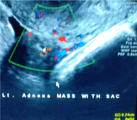
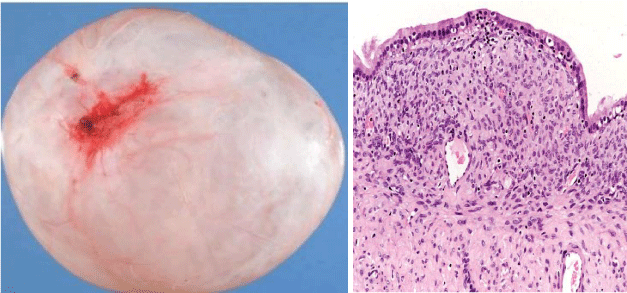
The patient was taken for ultra sonography .Trans abdominal and transvaginal Sonography showed live intrauterine pregnancy with CRL 1.00 cm corresponding to 7 weeks 1 day. There was a heterogeneous mass in left adnexa measuring approximately 4.3x3.9 cm. The mass showed Gestational sac measuring approximately 0.2 cm corresponding to 4 weeks. Left ovary was visualised separately. Right adnexa showed complex multicystic lesion measuring approximately 6.1x 4.9 cm It showed septal vascularity However no obvious solid areas where noted within. Right ovary was not separately visualised. Preliminary investigations were done and patient was taken up for laparotomy in view of bilateral adnexal masses. Per operatively gross morphology was: Right ovarian complex cystic mass ovarian cystectomy was done. Other side salpingostomy was done. Patient’s post-operative period was uneventful. Histopathology report came out to be serous cystadenoma and left adnexal mass was ectopic pregnancy.
Case 3
A 29-year-old woman (gravida 1 para 0) presented to the emergency department with a complaint of sudden onset of right-sided lower abdominal pain in the setting of a recent positive home pregnancy test. Her last known menstrual period was 10 weeks days prior to her presentation. She had no history of pelvic inflammatory disease or fertility treatment. On physical examination, she was hemodynamically stable with a blood pressure of 110/60 mm Hg and heart rate of 82 beats/min. Abdominal examination demonstrated a distended abdomen with diffuse abdominal tenderness that was maximal in the right iliac fossa with rebound tenderness and signs of peritonism. Cervical tenderness was elicited during bimanual examination, with cervical excitation maximal in the right adnexa. Per speculum examination demonstrated a long, closed, posterior cervical with no bleeding. Spontaneous pregnancy there was no history of induction of ovulation by drugs or artificial reproductive techniques. Transvaginal sonography demonstrated 3 gestational sacs-2 intrauterine and 1 right adnexal.
The crown rump lengths (CRL) of the twin intrauterine fetuses were
F1- 3.3 cm corresponding to gestations of 10 weeks and 2 days and
F2 - 0.8 cm corresponding to 6 weeks and 6 days, respectively
F1 shows normal cardiac activity
F2 shows absent cardiac activity with non-viable gestation.
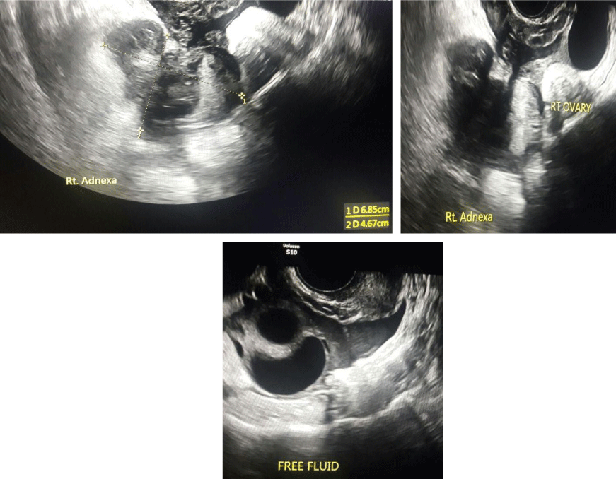
The right adnexa showed a mixed echogenic predominantly hyperechoic mass measuring approximately 6.8x 4.6 cm. Mass showed minimal vascularity.
Right ovary was visualised separately .Left ovary was visualised and appears normal. Ultrasonographic evidence of hemoperitoneum was present with a mild amount of free fluid within the pelvis. The patient underwent urgent laparoscopy that confirmed a ruptured right tubal ectopic pregnancy with hemoperitoneum. A laparoscopic right salpingectomy (using bipolar diathermy and scissors) was performed and effective hemostasis achieved. The histopathological examination of the excised right fallopian tube confirmed an ectopic pregnancy. The postoperative course was uneventful, with a hemoglobin level of 9.6 g/L, and the patient was discharged on postoperative day 5 on hematinics.
 Figure 1: Right adnexal mass with internal vascularity separate from right ovary. Irregular intrauterine gestational sac with Subchorionic hematoma.
 Figure 4: Trans vaginal Sonography shows heterotopic pregnancy, intrauterine pregnancy and left adnexal pregnancy. Left adnexal mass with gestational sac .Doppler shows vascularity of left adnexal mass.
Discussion
Duverney first described Heterotopic pregnancy in 1708.1,2. Presently, the use of Artificial reproductive techniques and fertility agents can increase a patient’s risk of Heterotopic pregnancy. This is possibly due to the combined effects of presence of tubal disease, high level of oestradiol and progesterone with hyper stimulation and the subsequent simultaneous transfer of several embryos into the uterus with retrograde flow into the fallopian tubes. [1,2]. Indeed, any factor predisposing a patient to an increased risk of ectopic pregnancy and/or multiple pregnancies can contribute to Heterotopic pregnancy [1,3,4,5]. In our patient (case 1), pregnancy also occurred in association with ovulation induction by Clomiphene citrate . Spontaneous heterotrophic pregnancy has an incidence of approximately 1 in 39,000. Most of the Heterotrophic pregnancy cases are diagnosed late, resulting in significant morbidity and occasional mortality. [6]. As no single investigation can predict the presence of a heterotopic pregnancy, it should be suspected in any patient, presenting with lower abdominal pain in the early phase of an obvious intrauterine pregnancy following fertility treatment. [1,7]. Often, abdominal and pelvic ultrasound fail to show an ectopic pregnancy or the ultrasound is misinterpreted because of the awareness of an existing intrauterine pregnancy [1,5]. Demonstration of an intrauterine pregnancy is not a reliable indicator for excluding an ectopic pregnancy [1,2]. Ectopic Pregnancy is normally visualised on ultrasound in three forms including “blob” sign, “bagel” (doughnut) sign or a gestational sac with a fetal pole [8]. But most ultrasound reports make no comment of a search for coexistent ectopic pregnancy when assessing an intrauterine gestation, because a heterotrophic pregnancy is still thought to be tremendously rare. For this reason, almost all ectopic pregnancies are diagnosed by excluding an intrauterine pregnancy [4]. Most commonly, the location of ectopic gestation in a heterotopic pregnancy is the fallopian tube. However, cervical and ovarian heterotopic pregnancies have also been reported [9,10]. Majority of the reported heterotopic pregnancies are of singleton intrauterine pregnancies. Triplet and quadruplet heterotopic pregnancies have also been reported, though extremely rare [11,12]. It can be multiple as well [13]. They can be seen frequently with assisted conceptions. Intrauterine gestation with hemorrhagic corpus luteum can simulate heterotopic/ectopic gestation both clinically and on ultrasound. Other surgical conditions of acute abdomen can also simulate heterotopic gestation clinically and hence the difficulty in clinical diagnosis. Bicornuate uterus with gestation in both cavities may also simulate a heterotopic pregnancy. High resolution transvaginal ultrasound with colour Doppler will be helpful as the trophoblastic tissue in the adnexa in a case of heterotopic pregnancy shows increased flow with significantly reduced resistance index. The illustrated case did not have any risk factor for the heterotopic gestation and presented with ruptured tubal pregnancy and history of p.v.bleeding. Patient did not suspected pregnancy as she had I.U.C.D insertion and was post partum last delivery 8 months back. Frequency of ovarian tumours being coexistent with pregnancy is 1:10007 and among these frequencies of being malignant is approximately 1:15000 to 1:32000 pregnancies. Most common ovarian masses encountered during pregnancy are functional cysts of ovary. The other ovarian masses in order are benign cystic teratomas, serous cyst adenomas, paraovarian cysts, mucinous cystadenomas and endometriomas. Benign serous tumours are unilocular (have one lobe); however if very large may be multilocular, contain clear fluid and have a smooth lining composed of columnar epithelial cells with cilia. On gross examination, the serous tumor may present as either a cystic lesion in which the papillary epithelium is contained within a few fibrous walled cysts, or the papillary projections may be away from the surface epithelium. It comprise 50% of all ovarian tumors. Sixty percent are benign (cystadenoma), 10% are borderline and 30% are malignant (cystadenocarcinoma). The benign serous tumors are most common in the third to fifth decades of life and may be 20-30 cm in size. Giant cysts are found in less than 1% of the cases of ovarian cysts with pregnancy +[14,5]. Torsion is the most common and serious complication of benign ovarian cysts during pregnancy. The other complications which might occur are rupture of cyst, infection, malignancy, impaction of cyst in pelvis, obstructed labour and malpresentations of fetus [15].
The gold standard in management of heterotopic pregnancies is surgery via laparoscopy or laparotomy, with the surgical approach guided by the clinical scenario. Laparoscopic approaches are preferred to open procedures except in cases of clinical shock with intra-abdominal haemorrhage where laparotomy may be the better suited procedure. In our case report, we opted for laparoscopic salpingectomy as the patient was hemodynamically stable and amenable to a laparoscopic versus an open surgical approach. Current evidence shows that despite our best efforts, the intrauterine component of a heterotopic pregnancy has a higher likelihood of miscarriage than sole intrauterine pregnancies, although survival rates of intrauterine heterotopic pregnancies have improved over the past few decades with those that proceed to live birth demonstrating no significantly different rates of adverse birth outcomes [16].
Conclusion
Heterotopic pregnancy is a potentially life-threatening condition that, while being rare, potentially has grave implications for both the mother and fetus. Ovarian tumors are rare in pregnancy and most common are functional cysts of pregnancy .High-risk groups warrant early pregnancy ultrasound as a part of routine antenatal care to enable early diagnosis and timely management. However, an absence of risk factors should not equate to exclusion. As highlighted in our case above, it must remain at the forefront of a clinician’s diagnostic algorithm in all women as it may occur in the absence of risk factors in a natural conception cycle. Despite it being a challenging diagnosis, clinical acumen along with skilled TVS and timely management is able to achieve optimal clinical outcomes.
References
- Strandell A, Thornburn J, Hamberger L. Risk factors for ectopic pregnancy in assisted reproduction. Fertil Steril. 1999; 71: 282-286. https://goo.gl/3bHCj6
- Li XH, Ouyang Y, Lu GX. Value of transvaginal sonography in diagnosing heterotopic pregnancy after in-vitro fertilisation with embryo transfer. Ultrasound Obset Gynecol. 2013; 41: 563-569. https://goo.gl/yKhGk2
- Marcus SF, Macnamee M, Brinsden P. Heterotopic pregnancies after in vitro fertilisation and embryo transfer. Hum Reprod. 1995; 10: 1232-1236. https://goo.gl/YKC5u6
- Ankum WM, Van der Veen F, Hamerlynck JV, Lammes FB. Transvaginal sonography and human chorionic gonadotrophin measurements in suspected ectopic pregnancy: a detailed analysis of a diagnostic approach. Hum Reprod. 1993; 8: 1307-1311. https://goo.gl/VZqR2g
- Barut A, Arikan I, Barut F, Harma M, Harma MI, Payasli B. Ovarian cancer during pregnancy. J Pak Med Assoc 61: 914-916. https://goo.gl/XdTq2b
- Ankum WM, Van der Veen F, Hamerlynck JV, Lammes FB. Transvaginal sonography and human chorionic gonadotrophin measurements in suspected ectopic pregnancy: a detailed analysis of a diagnostic approach. Hum Reprod. 1993; 8: 1307-1311. https://goo.gl/wVdjrM
- Fernandez H, Lelaidier C, Doumerc S, Fournet P, Olivenne SF, Frydman R. Nonsurgical treatment of heterotopic pregnancy: a report of six cases. Fertil Steril. 1993; 60: 428-432. https://goo.gl/BYySdB
- Barrenetxea G, Barinaga-Rementeria L, Lopez de, Larruzea A, Agirregoikoa JA, Mandiola M, et al. Heterotopic pregnancy: two cases and a comparative review. Fertil Steril. 2007; 87: 415-417. https://goo.gl/HnTB4Y
- Habana A, Dokras A, Giraldo JL, Jones EE. Cornual heterotopic pregnancy: contemporary management options. Am J Obstet Gynecol. 2000; 182: 1264-1270. https://goo.gl/mgn7yd
- Govindarajan MJ, Rajan R. Heterotopic pregnancy in natural conception. J Hum Reprod Sci. 2008; 1: 37-38. https://goo.gl/2GBcUH
- Simsek T, Dogan A, Simsek M, Pestereli E. Heterotopic triplet pregnancy (twin tubal) in a natural cycle with tubal rupture: case report and review of literature. J Obstet Gynaecol Res. 2008; 34: 759-762. https://goo.gl/zS5rm2
- Inion I, Gerris J, Joostens M, Vree BD, Kockx M, Verdonk P. An unsuspected triplet heterotopic pregnancy after replacement of two embryos. Hum Reprod. 1998; 13: 1999-2001. https://goo.gl/z8gDDc
- Jerrard D, Tso E, Salik R, Barish RA. Unsuspected heterotopic pregnancy in a woman without risk factors. Am J Emerg Med. 1992; 10: 58-60. https://goo.gl/xq12zx
- Palmer J, Jivraj S, Galimberti A, Paterson M. Serous ovarian carcinoma in pregnancy. BMJ Case Rep. 2009. https://goo.gl/f9RFes
- Umranikar S1, Umranikar A, Rafi J, Bawden P, Umranikar S, O’Sullivan B, et al. Acute presentation of a heterotopic pregnancy following spontaneous conception: a case report. Cases J. 2009; 2: 9369. https://goo.gl/o4P31E
- Bugatto F, Quintero-Prado R, Kirk-Grohar J, Melero-Jimenez V, Hervias-Vivancos B, Bartha JL. Heterotopic triplets: tubal ectopic and twin intrauterine pregnancy. A review of obstetric outcomes with a case report. Arch Gynecol Obstet. 2010; 282: 601-606. https://goo.gl/VUamBe
Authors submit all Proposals and manuscripts via Electronic Form!


