Case Report
T Lymphoblastic Transformation of Chronic Myeloid Leukemia with Isolated Maturing Precursors of Neutrophil Cell Line in the Cerebrospinal Fluid
Natasa Colovic1,2*, Danijela Lekovic1,2, Marko Jankovic3, Marija F. Dencic2 and Mirjana Gotic1,2
1Medical Faculty, University of Belgrade, Dr Subotica 8, 11000 Belgrade, Serbia
2Clinic of Hematology, Clinical Center of Serbia, Koste Todorovica 2, 11000 Belgrade, Serbia
3Institute of Microbiology and Immunology, Medical Faculty, University of Belgrade, Dr Subotica 1, Serbia
*Address for Correspondence: Natasa Colovic, Faculty of Medicine, University of Belgrade, Dr Subotica 8, 11010 Belgrade, Serbia, Tel: +381-11-361 55 69; E-mail: natasacolovic73@gmail.com
Dates: Submitted: 29 June, 2017; Approved: 11 July, 2017; Published: 13 July, 2017
Citation this article: Colovic N, Lekovic D, Jankovic M, Dencic MF, Gotic M. T Lymphoblastic Transformation of Chronic Myeloid Leukemia with Isolated Maturing Precursors of Neutrophil Cell Line in the Cerebrospinal Fluid. Int J Case Rep Short Rev. 2017;3(2): 027-030.
Copyright: © 2017 Colovic N, et al. This is an open access article distributed under the Creative Commons Attribution License, which permits unrestricted use, distribution, and reproduction in any medium, provided the original work is properly cited.
Keywords: Chronic myeloid leukemia; T lymphoblastic transformation; Imatinib mesylate; Neuroleukemia
Abstract
Introduction: Imatinib mesylate, a potent inhibitor of BCR/ABL tyrosine kinase, is the first therapeutic choice in Chronic Myeloid Leukemia (CML). It poorly penetrates the brain-blood barrier so that therapeutic concentrations in the Cerebrospinal Fluid (CSF) can hardly be achieved. In effect, rare clonogenic stem cells that had anchored in the Central Nervous System (CNS) may eventually proceed towards the blastic transformation there.
Case Report: We report on a 25 - yr - old male treated with imatinib mesylate who developed T lymphoblastic blast crisis without the clinical signs of neuroleukemia. After treatment with chemotherapy and preventive intrathecal methotrexate, cytosine-arabinoside and hydrocortisone he achieved the remission. Six months later he contracted a general relapse in the bone marrow and peripheral blood. Remarkably, his CSF cytology demonstrated only the cells representative of chronic phase of disease. The subclone raising the extensive blasts crisis was resistant to chemotherapy.
Conclusion: The CML relapsing in the CSF with a chronic phase clone in parallel with a T-lymphoblastic crisis in the bone marrow and the peripheral blood has never been reported so far. We emphasize the importance of CNS surveillance strategy using PCR methods in patients with signs of CNS involvement in CML.
Introduction
Central Nervous System (CNS) is rather infrequently affected in Chronic Myeloid Leukemia (CML) [1,2]. In the lymphoblastic transformation of CML the incidence of CNS involvment is comparable to that in Acute Lymphoblastic Leukemia (ALL) [1,2]. To the best of our knowledge, the relapse of Chronic Myeloid Leukemia in cerebrospinal fluid with a chronic phase clone coinciding with T lymphoblastic crisis in bone marrow and peripheral blood has not been previously reported. Here we report the clinical course in such a unique case.
Case Report
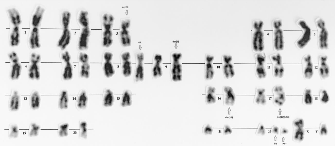
A 25 - yr - old male with a history of Philadelphia chromosome positive CML in chronic phase diagnosed on August 13th, 2013. At the time of diagnosis, he had a Sokal score of 1.08 and Hasford score of 207. The EUTOS score was 7, indicating low risk. The bone marrow aspirate was hypercellular and consistent with the diagnosis of chronic phase of CML. Cytogenetic analysis: 46, XY, t(3; 9; 16; 22)(p?;q34;q?;q11) (Figure 1). The PCR study detected the b3 a2 transcript. He was initially treated with Hydroxycarbamide, continuing the treatment afterwards with imatinib mesylate 400 mg/ p.d. In June 2014, on physical examination inguinal and axillar lymph nodes were enlarged with scrotal oedema and an oedematous upper part of the left leg. Histology of the inguinal lymph node showed a T lymphoblastic infiltration suggestive of the blastic transformation of CML. The hypercellular bone marrow aspirate was infiltrated with 62% of blasts which on flow cytometry showed the immunophenotype distinctive of T lymphoblasts: CD38, cCD3, CD2, CD7, CD13, CD33, CD11b+ and (mCD3, CD1a, nTdT, CD34, CD10-). Cytogenetic finding: 46, XY, t(3;9;16;22) (2)/ 47,XY,t(9;22) (1)/ 46, XY (5) (Figure 2). Flow cytometry of the cerebrospinal fluid was acellular at the time. He was treated with the Hyper-CVAD protocol and six preemptive cycles of methotrexate, cytosine-arabinoside and hydrocortisone intrathecally. He achieved a remission. The unrelated bone marrow transplantation was planned as the matching donor was found. Regrettably, the transplantation was postponed on patient’s demand and then on two occassions on demand of a donor. The patient was readmitted on June 16, 2015 presenting with a headache and signs of relapse. The blood count was as follows: Hb 95 g/ l, WBC 222 x 109/ l, and platelets 431 x 109/ l. In the differential the blasts were 9%, promyelocytes 2%, myelocytes 45%, metamyelocytes 17%, eosinophils 1%, basophyls 2%, lymphocytes 4%, and monocytes 2% and the bone marrow aspirate contained 5% of blasts. He was again treated with Hyper-CVADprotocol. Symptoms and signs of CNS leukemia (severe piercing headaches, dyplopia) persevered. Lumbar puncture was performed and the CSF retrieved contained 100 cells/µL. Only the maturing precursors of neutrophil cell line were detected including promyelocytes (CD66b - CD16- CD10-, ~12%), myelocytes (CD66+ CD16+, CD10-, ~71%) and metamyelocytes (CD66b+ CD16+, CD10+, ~7%) (Figure 3). The PCR study evidenced only the initial b3 a2 transcript. The absence of cells with a morphology and immunophenotype of blasts was quite impressive. The patient was treated with six intrathecal cytoablative courses of cytosar 100 mg and methotrexate 12 mg. Before the last intrathecal instilation there were still residual maturing cells of the neutrophil series present in the cerebrospinal fluid indicating lack of sensitivity to applied medication. His clinical condition aggravated and he passed away in October 2015.
Discussion
CML is a clonal myeloroliferative disease chracterized by proliferation and accumulation of mature haemopoietic cells in the peripheral blood and bone marrow occasionally accompanied by extramedullary haematopoiesis. The genetic molecular basis of the disease is fusion of the bcr gene on chromosome 22 with the abl gene on chromsome 9, forming the Philadelphia chromosome. Imatinib mesylate is the treatment of choice in the chronic phase of disease. It selectively inhibits the BCR/ABL kinase.
In about 5-10% of cases, variant translocations have been reported involving other chromosomes, but the involvement of chromosome 16 has been reported in ten cases only [3]. The majority of these reports are related to the cytogenetic finding and only few of them describe a respone on the treatment [4]. In preimatinib era it was generally accepted that the patients with variant Ph chromosomes have shorter survival [4]. However, after introduction of imatinib in the treatment of CML, patients with variant translocations showed similar survival and response to the patients with classic Ph chromosome [5]. Still, there are occasional reports of less favorable response on imatinib mesylate in patients with variant translocations due to the genomic instability [6].
The patient presented here was in a chronic phase of CML when treatment with hydroxyurea and imatinib mesylate was commenced. He responded cytologically but without acheiving a cytogenetic response. His clinical course suggested the resistance to imatinib therapy. After ten months he developed T lymphoblastic blast crisis and was treated with Hyper-CVAD protocol and a preventive intrathecal administration of six cycles of intrathecal methotrexate, cytosine-arabinoside and hydrocortisone. These resulted in a remission and the allogeneic bone marrow transplantation was planned. At that time there were no clinical or laboratory signs of CNS involvement. However, the patient’s condition deteriorated in June 2015. He presented with signs of neuroleukemia and a systemic disease relapse with high WBC count and karyotype evolution. Examination of the CSF showed presence of maturing precursors of neutrophil cell line cells distinctive of chronic phase CML which was confirmed by flowcytometry. The intrathecal treatment with cytosine-arabinoside, methotrexate and hydrocortisone was ineffective and the signs of CSF disease persisted.
Clinical evolution of CML in our patient was unusual because he developed a vigorous T lymphoblastic transformation which had initially responded well to chemotherapy. Further on in the course of disease he developed a relapsing blastemia in the peripheral blood followed by a leptomeningeal manifestation of the chronic phase subclone. The presence of blasts in the cerebrospinal fluid was never verified.
Whereas imatinib mesylate is most efficatious in the treatment of chronic phase CML, its efficacy in cases of lymphoblastic infiltration of CNS in blast crisis has not been evaluated extensively due to rarity of this complication. Penetration of the drug into the central nervous system is poor so the drug level is inadequate for killing leukemic stem cells, if any. In consequence, CNS may turn into a refuge for leukemic stem cells that had settled there [7-10]. Anecdotal reports suggest that imatinib does not penetrate the blood-brain barrier and has limited effects against leukemic blasts in the CSF. Low levels of the drug in cerebrospinal fluid have been documented in experimental animals and in humans [7-11]. Bornhaus, et al. [8] reported a patient with CML on imatinib who developed an isolated CNS relapse, although he achieved a complete cytogenetic response, as the patient had significantly lower level of imatinib in CSF then in blood. Similar observation was reported by Takayama in patient with Ph positive ALL and concurrent CNS and marrow relapse [7]. Isobe, et al. [12] reported isolated lymphoid blast crisis in CNS in a patient with CML after viral meningitis while he was in complete cytogenetic remission on imatinib therapy. Park, et al. [13], found 15 published cases of the isolated CNS relapse in CML patients treated with imatinib, 10 of which had lymphoblastic immunophenotype, 1 mixed lineage and 3 myeloid, while in 2 patientes the phenotype was not determined. Nine out of 15 had a full cytogenetic response. The median time from starting with imatinib ranged from 4 to 58 months and no predicting factors for CNS relapse were identified. So, the explanation for isolated CNS relapse could be the low level of the drug in CSF as a result of the poor CNS penetration mediated through P-glycoprotein efflux [14]. This emphasizes the problem of imatinib pharmacokinetics in the CNS, which might be worth to consider in patients taking the drug [13]. Even with a second generation of tyrosine-kinase inhibitor (dasatinib) that have an improved penetration into the CNS a single case of isolated CNS blast crises was reported [15].
Bornhauser, et al. [8] have suggested that the course of patients with CML on imatinib may be complicated by an unforeseen blast crisis in the CNS.
This quite unusual clinical course of CML, so far unreported, points at a need to define surveillance strategy with rare central and peripheral nervous system relapses associated with the blastic phase of CML. The analysis of BCR-ABL in cerebrospinal fluid and FISH analysis should be performed on the first sign of haematological relapse of CML, although regular CSF examination or prophylactic intratechal chemotherapy due to rarity of CNS involvement is not still indicated. No treatment guidelines for documented CNS relapse in this kind of patients have yet been defined.
Conflicts of Interest
The authors disclose no conflict of interests.
Funding
This work was supported by grant No. III 41004, Ministry of Education and Science, Republic of Serbia.
References
- Cortes J. Central nervous system involvement in adult lymphocytic leukemia. Hematol Oncol Clin North Am. 2001; 15: 145-62. https://goo.gl/n9FvZS
- Pfeifer H, Wassmann B, Hofmann WK, Komor M, Scheuring U, Bruck P, et al. Risk and prognosis of central nervous system leukemia in patients with Philadelphia chromosome-positive acute leukemias treated with imatinib mesylate. Clin Cancer Res. 2003; 9: 4674-81. https://goo.gl/zjrGFD
- Espinoza JP, Cardenas VJ, Jiménez EA, Angulo MG, Flores MA, García JR. A complex translocation (9; 22; 16)(q34;q11.2;p13) in chronic myelocytic leukemia. Cancer Genet Cytogenet. 2005; 157: 175-7. https://goo.gl/xky9mt
- Reid AG, Huntly BJ, Grace C, Green AR, Nacheva EP. Survival implications of molecular heterogeneity in variant Philadelphia-positive chronic myeloid leukaemia. Br J Haematol. 2003; 121: 419-27. https://goo.gl/V1KGDq
- Marzocchi G, Castagnetti F, Luatti S, Baldazzi C, Stacchini M, Gugliotta G. Variant Philadelphia translocations: molecular-cytogenetic characterization and prognostic influence on frontline imatinib therapy, a GIMEMA Working Party on CML analysis. Blood. 2011; 117: 6793-800. https://goo.gl/24xbYq
- Stagno F, Vigneri P, Del Fabro V, Stella S, Cupri A, Massimino M, Consoli C et al. Influence of complex variant chromosomal translocations in chronic myeloid leukemia patients treated with tyrosine kinase inhibitors. Acta Oncol. 2010; 49: 506-8. https://goo.gl/Cqkhj9
- Takayama N, Sato N, O'Brien SG, Ikeda Y, Okamoto S. Imatinib mesylate has limited activity against the central nervous system involvement of Philadelphia chromosome-positive acute lymphoblastic leukemia due to a poor penetration into cerebrospinal fluid. Br J Haematol. 2002; 119: 106-8. https://goo.gl/UMw1se
- Bornhauser M, Jenke A, Freriberg-Richter J, Radke J, Schuler US, Mohr B, Ehninger G, et al. CNS blast crisis of chronic myelogenous leukemia in a patient with a major cytogenetic response in bone marrow associated with low levels of imatinibmesylate and its N-desmethylated metabolite in cerebral spinal fluid. Ann Hematol. 2004; 83: 401-2. https://goo.gl/Ck87KZ
- Wolff NC, Richardson JA, Egorin M, Ilaria RL Jr. The CNS is a sanctuary for leukemic cells in mice receiving imatinibmesylate for Bcr/Abl-induced leukemia. Blood. 2003; 101: 5010-3. https://goo.gl/mCqwk1
- Rajappa S, Uppin SG, Raghunadharao D, Rao IS, Surath A. Isolated central nervous system blast crisis in chronic myeloid leukemia. HematolOncol. 2004; 22: 179-81. https://goo.gl/2Jzj72
- Altintas A, Cil T, Kilinc I, Kaplan MA, Ayyildiz O. Central nervous system blastic crisis in chronic myeloid leukemia on imatinibmesylate therapy: a case report. J Neuro-Oncol. 2007; 84: 103–05. https://goo.gl/pWUurq
- Isobe Y, Sugimoto K, Masuda A, Hamano Y, Oshimi K. Central nervous system is a sanctuary site for chronic myelogenousleukaemia treated with imatinibmesylate. Intern Med J. 2009; 39: 408–11. https://goo.gl/utoVZv
- Park MJ, Park PW, Seo YH, Kim KH, Seo JY, Jeong JH, Kim MJ, et al. A case of isolated lymphoblastic relapse of the central nervous system in a patient with chronic myelogenous leukemia treated with imatinib. Ann Lab Med. 2014; 34: 247-51. https://goo.gl/EEq6T2
- Dai H, Marbach P, Lemaire M, Hayes M, Elmquist WF. Distribution of STI-571 in the brain is limited by P-glycoprotein-mediated efflux. J Pharmacol Exp Ther. 2003; 304: 1085-92. https://goo.gl/fbB8jP
- Aftimos P, Nasr F. Isolated CNS lymphoid blast crisis in a patient with imatinib-resistant chronic myelogenous leukemia: case report and review of the literature. Leuk Res. 2009; 33: 178-180. https://goo.gl/X5S1ZU
Authors submit all Proposals and manuscripts via Electronic Form!


