Manouchehr Aghajanzadeh*, Alirza Jafanegad, Ali alive MD, Omid mosafaii, Yasaman Safarpoureand Samman Ayobi
Manouchehr Aghajanzadeh1*, Alirza Jafanegad2, Ali alive MD2, Omid mosafaii3, Yasaman Safarpoure3and Samman Ayobi3
1Department of thoracic surgery, Guilan University of Medical Sciences, Rasht, Iran
2Department of pulmonology, Guilan University of Medical Sciences, Rasht, Iran
3Respiratory diseases and TB Research Center, Razi Hospital, Guilan University of Medical Sciences, Rasht, Iran
*Address for Correspondence: Manouchehr Aghajanzadeh, Department of thoracic surgery, Guilan University of Medical Sciences, Rasht, Iran
Dates: Submitted: 01 January, 2017; Approved: 17 February, 2017; Published: 21 February, 2017
Citation this article: Aghajanzadeh M, Jafanegad A, Alive A, Mosafaii O, Safarpoure Y, et al. Repot of Huge Primary Mediastinal Carcinoid Tumors with Horsiness. Int J Case Rep Short Rev. 2017;3(2): 016-018
Copyright: © 2017 Aghajanzadeh M, et al. This is an open access article distributed under the Creative Commons Attribution License, which permits unrestricted use, distribution, and reproduction in any medium, provided the original work is properly cited.
Abstract
A carcinoid tumor can occur in a variety of sites, including the mediastinum. Carcinoid tumor arising from the mediastinum is invariably related to the thymus. Isolated origin of mediastinal carcinoids is rare, especially in the posterior mediastinum. Only two cases of posterior mediastinal carcinoids have been reported so far. These were assumed to be arising from ectopic thymus tissue. We report a case of a 48-year-old man who presented with dyspnoea, horsiness and dry cough due to giant carcinoid tumor of the posterior mediastinum, the pedicle originating from the posterior mediastinum, not related to the thymus. She underwent thoracotomy and resection that provided relief. The Microscopic and immunochemical studies revealed extrathymic and show more pronounced cytologic atypia, increased mitotic activity (approximately 3-/ 410 HPF), Grade II) Grade II or (intermediate grade neuroendocrine neoplasm (a typical carcinoid).
Introduction
Mediastinal masses present challenging problems in thoracic surgery. One of these tumors is neuroendocrine carcinoma Carcinoid tumor can occur in a variety of sites, such as the lung, thymus, parathyroid, ovary, and biliary and gastrointestinal tracts mediastinum [1,2]. Occasionally, the tumor arises from nonparenchymal soft tissue, including the mesentery, retroperitoneum, inferior vena cava, or the presacral region [3,4]. 3-8 in revives of literature, we found only two cases of posterior mediastinal carcinoids have been reported so far [1-4]. Most of them remain asymptomatic for long and by the time the pressure symptoms as a cough, dyspnea and chest pain develop, in this time tumors are quite advanced. Herein, we present a case of neuroendocrine carcinoma of the posterior mediastinum with a cough, dyspnea, weight loss and horsiness and discuss its symptoms, diagnosis tools, treatment and pathologic implications this case of primary neuroendocrine carcinoma of posterior mediastinum.
Case Report
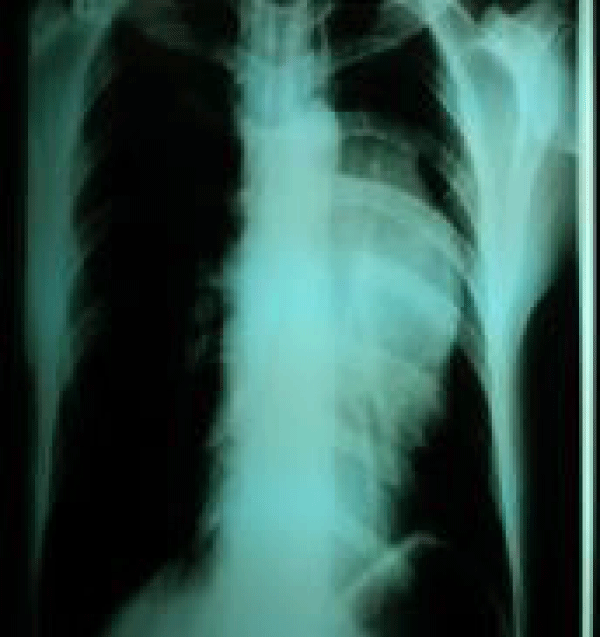
A 48-year-old man presented with a dry cough and dyspnea since six months that was associated with weight loss (10 Kg) and change in voice. Wheezing was hearted on the left chest. Cervical region was normal chest radiograph (posterio-anterior view) (Figure 1) revealed a large left para-cardiac and aorta mass, that was confirmed to be a mediastinal mass (20cm x 16cm x 14cm) on computed tomography, revealing huge mass with areas of necrosis and compress on the left pulmonary and bronchus (Figure 2) beta HCG, LDH, alfa FP and CEA normal. The patient has referred for CT-scan guided trans-thoracic-needle biopsy, the pathologist report was neuroendocrine (carcinoid) tumor. The patient was undergone to a left posterior-lateral thoracotomy. The tumor was found to occupy most of the left hemi thorax extending from posterior to anterior mediastinum. It was resected completely along with pedicle that was arising from the left para vertebral area between the left pulmonary artery and the arch of the aorta. The lesion was lobulated, well-encapsulated except over the region of the pedicle it was 14cm x 11cm x 10cm in size and weighed 850 gm and the cut surface showed solid grey-white and the tumor was encapsulated but fragile (Figure 3). Microscopic findings showed neoplastic tissue composed of nests of the monotonous small cuboidal cell with round nuclei & inconspicuous multiple nucleoli surrounded by delicate fibrovascular bundles. More pronounced cytologic atypia, increased mitotic activity (3-4/ 10 hpf) figures but no tumor necrosis is seen. The adipose tissue contains a reactive lymph node (intermediate grade neuroendocrine neoplasm (a typical carcinoid, Grade II). The post-operative period was uneventful. Horsiness was improved the patient is being followed-up and is doing well at the end of six months and weight gained 5 Kg.
Figure 2
CT chest showing a large non-enhancing cystic lesion in the mediastinum with hypodense and hyperdense areas and origin of pedicle in the posterior mediastinum.

Discussion
The carcinoids are a neuroendocrine group of tumors, arising from Kulchitsky cells. They can arise from along the gastrointestinal tract or the bronchial mucosa. The tumor can Release serotonin may produce carcinoid syndrome (flushing, diarrhea, and wheezing). Carcinoid was defined in 1907 by Oberndorfer but is now more relevantly named as the neuroendocrine tumor [2]. The origin of neuroendocrine tumors of the mediastinum is either from thymus or from the ectopic neuroectodermal tissue. Rosai and Higa are credited with the first description of such tumors in the thymic region [5]. There is the marginal relevance of the precise distinction between thymic and mediastinal carcinoids [6]. However, few cases of posterior mediastinal carcinoid tumor reported so far have been attributed to ectopic thymic tissue [7]. Clinically, patients may be asymptomatic or may present with symptoms of compression of the mediastinal structures [7]. As but our patient present with dry a cough and dyspnea since six months that was associated with weight loss (10 Kg) and change in voice with the giant mass of mediastinum. Less frequently, a carcinoid tumor may present with of various endocrine disorder as Cushing syndrome [2].
Only few reports have proposed it as a primary carcinoid tumor arising from neuroectodermal elements of mediastinum similar to the primary carcinoid [8]. However, no previous study has been conducted on primary neuroendocrine carcinoma arising from the posterior mediastinum, [1] while several cases of primary neuroendocrine carcinoma and carcinoid arising from the mesentery, retroperitoneum, inferior vena cava, and the presacral region have been reported [3-4]. In our case, the tumor was to occupy the two third of the left hemithorax. Origin the pedicle of the tumor was arising from the left paravertebral area between the left pulmonary artery and the arch of the aorta. In our case, the tumor was distinctly separate from thymus and there was no evidence of thymic cells in the specimen. There was no evidence of regional lymph node. The differential diagnosis of neuroectodermal lesion included paraganglioma, lymphoma neuroblastoma, peripheral neuroectodermal tumor and peripheral neuroepithelioma. The immunohistochemical findings excluded these lesions from neuroectodermal tumor [1,2,8,9]. Based on histology and immunohistochemistry, Carcinoid tumors divided to three type or grade [1,2,8,9]: Grade I or well-differentiated neuroendocrine tumor is positive for neuroendocrine cells (synaptophysin and chromogranin) with a low proliferative index (K-67 being 2% to 3%).Grade II or moderately differentiated usually show more pronounced cytologic atypia, increased mitotic activity (approximately 4-9/ 10 HPF), Grade III (poorly differentiated or small cell carcinoma)based on histology and degree of differentiation (monotonous cells with bland morphology and inconspicuous nucleoli and small foci of necrosis) and more frequent and extensive areas of necrosis and vascular invasion while poorly differentiated neuroendocrine carcinomas show marked cytologic atypia very high mitotic activity (> 10 MF / 10HPF), extensive areas of necrosis and frequent foci of vascular invasion [1,2,8,9].
Based on histology and immunohistochemistry, the present tumor is grade II (moderately differentiated or intermediate or atypical carcinoid moderately differentiated neuroendocrine. The patient is being followed-up and is doing well at the end of six months and weight gained of 5 Kg.
Figure 3
CT chest showing a large non-enhancing cystic lesion in the mediastinum with hypodense and hyperdense areas and origin of pedicle in the posterior mediastinum.

References
- yasushi horie, masako kato. neuroendocrine carcinoma of the posterior mediastinum a possible primary lesion. Arch Pathol Lab Med. 1999; 123: 933-936.
- sunil joshi, arunkumar haridas, priti lata rout. primary carcinoid of posterior mediastinum: truth or myth!. Indian J Chest Dis Allied Sci. 2010; 52: 241-243.
- Barnardo DE, Stavrou M, Bourne R, Bogomoletz WV. Primary carcinoid tumor of the mesentery. Hum Pathol. 1984; 15: 796-798.
- Horenstein MG, Erlandson RA, Gonzalez-Cueto DM, Rosai J. Presacral carcinoid tumors: report of three cases and review of the literature. Am J Surg Pathol. 1998; 22: 251-255.
- Rosai J, Higa E. Mediastinal endocrine neoplasm of probable thymic origin related to carcinoid tumour: clinicopathologic study of 8 cases. Cancer. 1972; 29: 1061-1074.
- Moran CA, Suster S. Thymic neuroendocrine carcinomas with combined features ranging from well-differentiated (carcinoid) to small cell carcinoma: a clinicopathologic and immunohistochemical study of 11 cases. Am J Clin Pathol. 2000; 113: 345-350.
- Cardillo G, Treggiari S, Paul MA, De Massimi AR, Remotti D, Graziano P, et al. Primary neuroendocrine tumours of the thymus: a clinicopathologic and prognostic study in 19 patients. Eur J Cardiothorac Surg. 2010; 37: 814-818.
- Renshaw AA, Haja JC, Neal MH, Wilbur DC; Cytopathology Resource Committee, College of American Pathologists. Distinguishing carcinoid tumor of the mediastinum from thymoma: correlating cytologic features and performance in the College of American Pathologists Interlaboratory Comparison Program in Nongynecologic Cytopathology. Arch Pathol Lab Med. 2006; 130: 1612-1615.
Authors submit all Proposals and manuscripts via Electronic Form!


