Shwetabh Sinha and Tabassum Wadasadawala*
1Department of Radiation Oncology, Advanced Centre for Treatment Research and Education in Cancer (ACTREC), Tata Memorial Centre, Kharghar, Navi Mumbai, India
*Address for Correspondence: Tabassum Wadasadawala, Associate Professor, Department of Radiation Oncology, Advanced Centre for Treatment Research and Education in Cancer (ACTREC), Tata Memorial Centre, Kharghar, Navi Mumbai
Dates: Submitted: 14 March 2016; Approved: 06 April 2017; Published: 10 April 2017
Citation this article: Sinha S, Wadasadawala T. A Very Rare Case of Malignant Pilomatrix Carcinoma of the Mammary Skin. Int J Cancer Cell Biol Res. 2017; 2(1): 006-008
Copyright: © 2017 Sinha S, et al. This is an open access article distributed under the Creative Commons Attribution License, which permits unrestricted use, distribution, and reproduction in any medium, provided the original work is properly cited.
Introduction
Pilomatrix carcinoma is extremely rare malignant tumours arising from hair follicles with around 80 cases reported till date. Owing to the paucity of literature and similarity of clinic pathological features with its benign counterpart they are often misdiagnosed. These tumors are known to be locally aggressive with low albeit significant metastatic potential. Surgical excision is the mainstay of treatment with adjuvant radiotherapy being used historically for improving local control. We present a case of an extremely unusual site (breast) of pilomatrix carcinoma. To the best of our knowledge no case of pilomatrix carcinoma of the mammary skin has been reported till date.
Case Report
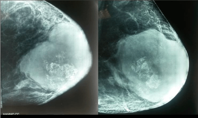
A 50 year old, postmenopausal lady presented to us in January 2015 with a history of left breast lump for 2 years for which she was treated outside. According to her the lump was pea sized to begin and it gradually grew to a size of tennis ball in 2 years. Fine Needle Aspiration Cytology (FNAC) was performed initially which was indeterminate. Subsequently, a core biopsy was performed which showed adnexal/metaplastic tumour which was negative for Estrogen (ER), Progesterone (PR) and Her2neu. On Mammography (MMG), a large lobulated opacity with chunky calcifications in the left breast was observed (BIRADS IV) (Figure 1). On UltraSonoGraphy (USG), a well-defined capsulated heteroechoic space occupying lesion with dense calcification of size 7.6 cm x 3.8 cm with surrounding hypochoic collection was seen. A baseline Positron Emission Tomography-Computerized Tomography (PET CT) scan showed a left breast primary of size 7.0 x 5.0 cm and SUVmax 16.3 with metabolically active and enlarged (size 1.3 cm, SUVmax 6.0) axillary lymph nodes. She received 3 cycles of Docetaxel and Epirubicin till December 2014 after which an interval PET CT showed a left breast lesion of 7.5 x 6 cm size and SUVmax of 10.3 and no active lymph nodes. She subsequently received one more cycle of chemotherapy (Docetaxel and Cisplatin) after which she presented to us. The reason for change in the chemotherapy was not documented but could be related to disease progression as she presented to us with a large 15x15 cm hard, non-tender lump in the left breast involving all the quadrants of the breast. The lump was fixed to underlying skin and chest wall. A soft left axillary lymph node was also felt while the rest of local examination was clear. The outside core biopsy review showed scanty carcinoma of intermediate nuclear grade. Differential diagnosis of carcinoma of skin adnexal origin, non keratinizing squamous cell carcinoma and metaplastic carcinoma was considered. She underwent left modified radical mastectomy in view of large size and fixity to skin. The histopathology showed it to be a malignant pilomatrixoma of tumor size 15 cm x 15 cm x10 cm with invasive component and necrosis. All cut margins were free, no lymphovascular emboli or perineural invasion was noted and all the 27 nodes dissected were negative. She was planned for adjuvant treatment in view of malignant nature of the tumor. However, chemotherapy was not given as benefit was unlikely considering poor response to neoadjuvant chemotherapy. She was treated with adjuvant Radiotherapy (RT) to a dose of 50 Gy /25 # / 5 weeks to the left chest wall which she tolerated well and concluded in May 2015. She is currently on follow up and locally controlled for the past 24 months since the completion of radiotherapy.
Figure 1
Mammographic images of left breast showing large lobulated opacity with chunky calcifications.

Discussion
Pilomatrixomas have been known since 1880 when they were first described by Malharbe and Chenantanis. They constitute about 1% of all skin tumours. Erroneously thought of arising from sebaceous glands, now they are known to arise from hair follicle matrix.
Malignant pilomatrixomas or pilomatrix carcinoma was first described in 1980 by Lopansiri and Mihm and till date around 80 cases have been reported [1]. Most of these tumours were located in head and neck region followed in sequence by upper limbs, lower limbs and scalp [2]. A male preponderance (5:1) has been seen and these tumours are more commoner in age >50 years and white race [3,4]. Repeated trauma triggering an inflammatory response has been hypothesized as an etiological factor for benign pilomatrixomas. Pilomatrix carcinomas are also found more frequently in persons suffering from Gardner's syndrome and myotonic dystrophy [5,6].
Most pilomatrix carcinoma present as a hard, fixed, painless mass of varying size and history ranging from months to years, suggesting a benign to malignant transformation Pathologically, the biggest challenge lies in differentiating a benign growth from malignant carcinomas. The morphological features of carcinomas include a pT size > 4cm, basaloid cell proliferation, nuclear pleomorphism, atypical mitosis, number of mitosis > 16-20 per hpf, infiltration of skin and soft tissue and vascular or lymphatic infiltration [7,8]. No definitive criteria for differentiation exists as till date no IHC or flow-cytometric markers have been identified [3]. Lymph node metastasis has hardly been reported in literature. Wide local excision (excision with 1 or 2 cm margin) has been the modality of choice and a recurrence rate of up to 59% has been reported in 5-17 months. Adjuvant radiotherapy has been show to decrease the local recurrence rate in sub-sites like head and neck, extremities and scalp [9,10,11,7].
The dose that has been used in other sites is between 45-60 Gy using conventional fractionation. Chemotherapy (mostly in the form of 5-fluorouracil and Cisplatinum) has been used in head and neck and other sites, however they appear to have no benefit except in metastatic setting [1,12,13]. Metastasis has also been reported in 10 of the cases mainly to lung and rarely to liver, brain and other sites [14,15,13].
To the best of our knowledge occurrence of pilomatrix carcinoma of the breast has not been reported till date although its benign counterpart (pilomatrixoma) is a well-defined entity. The incidence of benign pilomatrixoma in breast is estimated to be around (1:100000) [16-18]. On MMG, breast pilomatrixomas appear as pleomorphic coarse irregular calcifications (BIRADS IV-V). On USG they appear as hypoechoic nodule with irregularity [19,20]. Treatment usually consists of wide local excision with excellent long term outcomes [21].
We gave adjuvant RT to this patient by extrapolating data from pilomatrix carcinoma from other sites. The dose of 50Gy / 25# seems adequate as the patient is locally controlled 24 months post RT.
Conclusion
Pilomatrix carcinomas are extremely rare tumours of the breast, which may simulate the much more common breast carcinomas of ductal and lobular origin. Clinically they can be difficult to diagnose but may be distinguished radiologically. It also requires a high degree of pathological expertise to differentiate from its benign counterpart. Adjuvant radiotherapy to following surgical excision seems to achieve adequate local control.
References
- Lopansri S, Mihm MC Jr. Pilomatrix carcinoma or calcifying epitheliocarcinoma of malherbe. A case report and review of literature. Cancer. 1980; 45: 2368-2373. https://goo.gl/19cYMG
- Liu CC, Hoy M, Matthews TW, Guggisberg K, Chandarana S. Pilomatrix carcinoma of the head and neck: Case report and literature review. Head Neck Oncol. 2014; 6: 12. https://goo.gl/NcVqvy
- Bremnes RM, Kvamme JM, Stalsberg H, Jacobsen EA. Pilomatrix carcinoma with multiple metastases: report of a case and review of the literature. Eur. J. Cancer. 1999; 35: 433-437. https://goo.gl/u7Vzjy
- Hardisson D, Linares MD, Cuevas-Santos J, Contreras F. Pilomatrix carcinoma: a clinicopathologic study of six cases and review of the literature. Am. J. Dermatopathol. 2001; 23: 394-401. https://goo.gl/Yz1zXj
- Pujol RM, Casanova JM, Egido R, Pujol J, de Moragas JM. Multiple Familial Pilomatricomas: A Cutaneous Marker for Gardner Syndrome? Pediatr. Dermatol. 1995; 12: 331-335. https://goo.gl/whZZBi
- Mueller CM, Hilbert JE, Martens W, Thornton CA, Moxley RT 3rd, Greene MH, et al. Hypothesis: neoplasms in myotonic dystrophy. Cancer Causes Control CCC20. 2009; 2009-2020. https://goo.gl/bqgvIR
- Sau P, Lupton GP, Graham JH. Pilomatrix carcinoma. Cancer. 1993; 71: 2491-2498. https://goo.gl/05bV3d
- Petit T, Grossin M, Lefort E, Lamarche F, Henin D. Pilomatrix carcinoma: histologic and immunohistochemical features. Two studies. Ann. Pathol. 2003; 23: 50-54. https://goo.gl/eOgbE9
- Aherne NJ, Fitzpatrick DA, Gibbons D, Armstrong JG. Pilomatrix carcinoma presenting as an extra axial mass: clinicopathological features. Diagn. Pathol. 2008; 3: 47. https://goo.gl/LGA3dH
- Bhasker S, Bajpai V, Bahl A, Kalyanakuppam S. Recurrent pilomatrix carcinoma of scalp treated by electron beam radiation therapy. Indian J. Cancer. 2010; 47: 217. https://goo.gl/pSwFow
- Yadia S, Randazzo CG, Malik S, Gressen E, Chasky M, Kenyon LC, et al. Pilomatrix Carcinoma of the Thoracic Spine: Case Report and Review of the Literature. J. Spinal Cord Med. 2010; 33: 272-277. https://goo.gl/Fecjxn
- Eluecque H, Gisquet H, Kitsiou C, Simon E, Chassagne JF, et al. Pilomatrix Carcinoma: A Case Report. J Clin Exp Dermatol Res. 2012; 3: 152. https://goo.gl/1T6Lzl
- Arslan D, Gunduz S, Avci F, Merdin A, Tatli AM, Uysal M, et al. Pilomatrix carcinoma of the scalp with pulmonary metastasis: A case report of a complete response to oral endoxan and etoposide. Oncol. Lett. 2014; 7: 1959-1961. https://goo.gl/wMPmvS
- Gould E, Kurzon R, Kowalczyk AP, Saldana M. Pilomatrix carcinoma with pulmonary metastasis. Report of a case. Cancer. 1984; 54: 370-372. https://goo.gl/BrsNVY
- Walker DM, Dowthwaite S, Cronin D, Molden-Hauer T, McMonagle B. Metastatic pilomatrix carcinoma: Not so rare after all? A case report and review of the literature. Ear. Nose. Throat J. 2016; 95: 117-120. https://goo.gl/X6jPZB
- Pascual A, Casado I, Colmenero I, Pelayo A, Asenjo JA. Fine needle aspiration cytology of pilomatrixoma of the breast. Acta Cytol. 2000; 44: 274-276. https://goo.gl/xNmo33
- Kim SJ. Pilomatricoma of the breast in an adolescent girl: sonographic findings. J. Med. Ultrason. 2013; 40: 149-151. https://goo.gl/PPXTvT
- Nori J, Abdulcadir D, Giannotti E, Calabrese M. Pilomatrixoma of the breast, a rare lesion simulating breast cancer: a case report. J. Radiol. Case Reports. 2013; 7: 43-50. https://goo.gl/PQJ9h8
- Gilles R, Guinebretiere JM, Gallay X, Vanel D. Pilomatrixoma mimicking male breast carcinoma on mammography. AJR Am. J. Roentgenol. 1993; 160: 895. https://goo.gl/tYjYT7
- Imperiale A, Calabrese M, Monetti F, Zandrino F. Calcified pilomatrixoma of the breast: mammographic and sonographic findings. Eur. Radiol. 2001; 11: 2465-2467. https://goo.gl/2XUo3J
- Guinot-Moya R, Valmaseda-Castellon E, Berini-Aytes L, Gay-Escoda C. Pilomatrixoma. Review of 205 cases. https://goo.gl/GBFR7w
Authors submit all Proposals and manuscripts via Electronic Form!


