Case Report
Postoperative Diagnosed Coronary Artery Spasm during Tooth Extraction under Intravenous Sedation
Kentaro Ouchi*3
Department of Dental Anaesthesiology, Field of Maxillofacial Diagnostic and Surgical Science; Faculty of Dental Science, Kyushu University, Japan
*Address for Correspondence: Kentaro Ouchi, Department of Dental Anesthesiology, Field of Maxillofacial Diagnostic and Surgical Sciences; Faculty of Dental Science, Kyushu University, 3-1-1 Maidashi, Higashi-ku, Fukuoka 812-8582, Japan
Dates: Submitted: 15 December 2016; Approved: 29 December 2016; Published: 02 January 2017
Citation this article: Ouchi K. Postoperative Diagnosed Coronary Artery Spasm during Tooth Extraction under Intravenous Sedation. American J Anesth Clin Res. 2017;3(1): 005-008
Copyright: © 2017 Ouchi K. This is an open access article distributed under the Creative Commons Attribution License, which permits unrestricted use, distribution, and reproduction in any medium, provided the original work is properly cited.
Keywords: Coronary artery spasm; Intraoperative managemen; Intravenous sedation
Abstract
This report describes a case of coronary artery spasm associated with ST-segment depression during intravenous sedation.
A 77-year-old woman with periapical periodontitis was scheduled for tooth extraction. She had a history of diabetes mellitus, but no history or evidence of ischemic heart disease. Intravenous propofol maintained sedation (Mackenzie and Grant sedation score 3-4). Four minutes after beginning the operation, ST-segment depression of 0.2mV was noted on the Electrocardiogram (ECG) monitor. Nitro-glycerine was immediately administered. Propofol administration was discontinued at the end of surgery as ST-segment depression showed a tendency to recover. On cessation of anesthesia, the patient had no anginal pain and ST-segment depression had recovered. Coronary angiography after anesthesia showed a normal coronary artery without significant narrowing.
As postoperative coronary angiography showed no abnormalities, coronary artery spasm was diagnosed as the cause of the ECG change. Possible inducing factors in the present case were diabetes mellitus and shallow anesthesia due to intravenous sedation. Careful anesthetic management is required to prevent intraoperative coronary artery spasm even in patients without a history of ischemic heart disease.
Introduction
The coronary artery spasm is the state that coronary arteries constrict transiently. The intraoperative coronary artery spasm has many reports from Japan [1]. Because, there is ethnic difference of genetic polymorphism relating to Nitric Oxide (NO) acting on coronary endodermis[2]. Slight coronary artery spasm may cause serious coronary artery spasm, besides may cause cardiac arrest [3-5].
This report describes a case of coronary artery spasm associated with ST-segment depression during intravenous sedation.
Case
A 77-year-old woman with periapical periodontitis was scheduled for tooth extraction. She had a history of diabetes mellitus, but no history or evidence of ischemic heart disease.
In first visit, patient presented with blood glucose level 240 mg/ dl and HbA1c 10.0 %. The patient started treatment of diabetes mellitus. In three month after first visit, patient presented with blood glucose level 170 mg/ dl and HbA1c 6.5 %. Accordingly, patient was scheduled for tooth extraction at three month after first visit. Preoperative ECG showed a normal sinus rhythm.
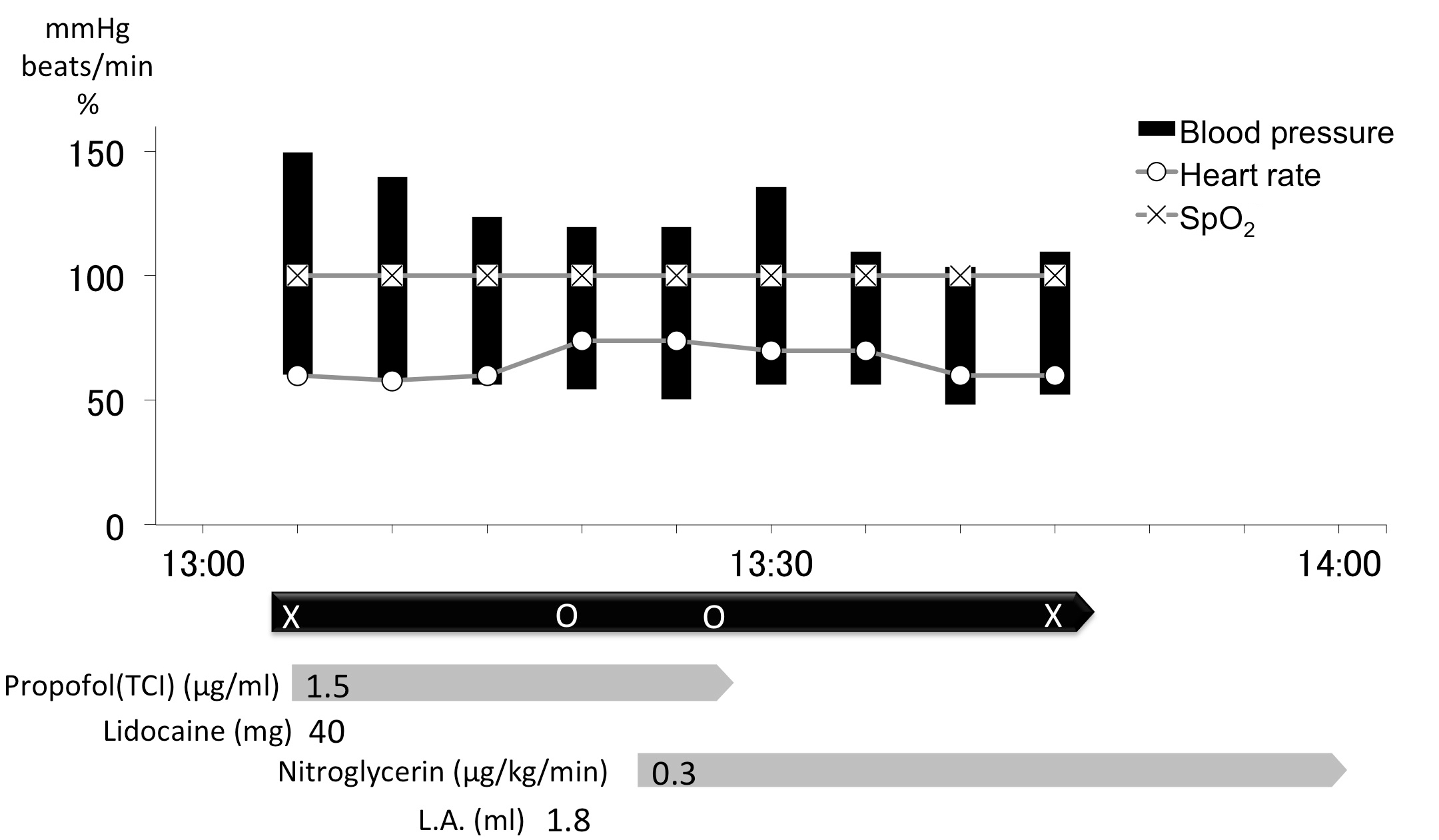
Anesthesiologist certified by the Japanese Board of Dental Anaesthesiologists' performed case of anesthesia sedation. Intravenous propofol inducted sedation, and maintained (Mackenzie and Grant sedation score 3-4) (Figure 1). Four minutes after beginning the operation, ST-segment depression of 0.2mV was noted on the electrocardiogram (ECG) monitor (Figure 2). Nitroglycerine was immediately administered. Propofol administration was discontinued at the end of surgery as ST-segment depression showed a tendency to recover. On cessation of anesthesia, the patient had no anginal pain and ST-segment depression had recovered (Figure 3).
Coronary angiography was performed several days later. Coronary angiography showed a normal coronary artery without significant narrowing.
Figure 1
Record of Anesthesia & Clinical Research Intravenous sedation was performed with propofol. After beginning operation, ST-segment depression on the ECG monitor followed was observed. Administration of the nitroglycerine was started immediately. Operation Time: 8 minutes; Anesthesia & Clinical Research Time: 37 minutes An abbreviated designation and symbol are meaning to the following matters. X: Anesthesia & Clinical Research start / finish O: Operation start / finish L.A.: Local anesthesia (infiltration anesthesia) Adrenaline include 2% Lidocaine

Figure 2
Intraoperative ECG in Lead II
ST segment decreased about 4 minutes after beginning of operation. ST-segment depression of 0.2mV on the ECG monitor followed was observed.

Discussion
In dental practice, intravenous anesthesia without tracheal intubation is often used for such as patients who are difficult to treat due to dental anxiety [6,7]. This report describes a case of coronary artery spasm associated with ST-segment depression during intravenous sedation.
ST-segment depression on ECG indicated myocardial ischemia due to supply-demand imbalance of myocardial oxygen [8]. As postoperative coronary angiography showed no abnormalities, coronary artery spasm was diagnosed as the cause of the ECG change. Although many cases of coronary artery spasm with ST-segment elevation have been reported, there have been few reports of ST-segment depression as in the present case [9-12]. ST-segment elevation reflects coronary occlusion causing transmural ischemia, while ST-segment depression reflects subendocardial ischemia causing coronary stenosis with developed collateral circulation or without occlusion [4]. If severe coronary spasm develops, ST-segment depression may shift to ST-segment elevation and severe arrhythmia, necessitating early administration of a coronary vasodilator [13,14]. Nitroglycerine used in the present case showed good effect [4].
Inducing factors of coronary artery spasm include diabetes mellitus, shallow anesthesia, vagal reflex, hypertension, older age, hyperventilation, smoking, and history of ischemic heart disease [9,15]. Shallow anesthesia is reported to cause coronary artery spasm due to imbalance in the automatic nervous system [16]. Our patient was in an optimal sedative state, but in terms of intravenous sedation, the sedation level was shallower than general anesthesia and it would appear, therefore, that intravenous sedation was the inducing factor of coronary artery spasm in our patient. Patients with diabetes are often asymptomatic for myocardial ischemia (Silent Myocardial Ischemia) and in those with severe diabetes; coronary artery spasm might occur asymptomatically [17]. Accordingly, even with no history or evidence of ischemic heart disease, such patients may develop electrocardiogram abnormalities requiring early detection and treatment.
An a-agonist, adrenaline is reported to cause coronary artery spasm [16]. In one of those reports, when patient with coronary artery spasm history received adrenaline 0.7 mg, ECG shows ST-segment elevation [18]. It reported that tachycardia (100 beats/ min) and hypertension (210 mmHg systolic blood pressure) were indicated. Most of coronary artery spasm patients have structural coronary stenosis [9-14]. Thus, it considered that that report is to cause myocardial demand supply balance worsening due to increase in cardiac work. On the other hand, in the present case, local anesthetics included adrenaline, but adrenaline dose was 0.0225 mg. Moreover, increase in cardiac work was not showed. Moreover, coronary angiography not showed significant narrowing. Thus, in the present case, it is not considered cause by adrenaline.
Possible inducing factors in the present case were diabetes mellitus and shallow anesthesia due to intravenous sedation. Careful anesthetic management is required to prevent intraoperative coronary artery spasm even in patients without a history of ischemic heart disease.
Conclusion
Coronary Artery Spasm with ST-segment depression occurred during intravenous sedation. Possible inducing factors in the present case were diabetes mellitus and shallow anesthesia due to intravenous sedation. Careful anesthetic management is required to prevent intraoperative coronary artery spasm even in patients without a history of ischemic heart disease.
Acknowledgments
This work was presented, in part, on July 29, 2011, at the 27th Regional Meeting of the Japanese Dental Society of Anesthesiology in the Chugoku and Shikoku Districts (Chairperson: Prof. Hiroshi Kitahata), Tokushima, Shikoku, Japan.
References
-
1. Pristipino C, Beltrame JF, Finocchiaro ML, Hattori R, Fujita M, Mongiardo R, et al. Major racial differences in coronary constrictor response between japanese and caucasians with recent myocardial infarction. Circulation. 2000; 101: 1102-1108.
2. Murase Y, Yamada Y, Hirashiki A, Ichihara S, Kanda H, Watarai M, et al. Genetic risk and gene-environment interaction in coronary artery spasm in Japanese men and women. Eur Heart J.2004; 25: 970-977.
3. Kawano H, Motoyama T, Yasue H, Hirai N, Waly HM, Kugiyama K, et al. Endothelial function fluctuates with diurnal variation in the frequency of ischemic episodes in patients with variant angina. J Am Coll Cardiol. 2002; 40: 266-270.
4. Hatae H. Clinical practice and mechanism of the coronary artery spasm. J Jpn Physicians Associ. 2006; 21: 184-189.
5. Fujii K. A case of intraoperative cardiac arrest. J Jpn Dent Soc Anesthesiol. 1993; 21: 123-130.
6. Ouchi K, Fujiwara S, Sugiyama K. Acoustic method respiratory rate monitoring is useful in patients under intravenous anesthesia. J Clin Monit Comput. 2016; 31: 59-65.
7. Ouchi K, Sugiyama K. Required propofol dose for anesthesia and time to emerge are affected by the use of antiepileptics: prospective cohort study. BMC Anesthesiol. 2015; 15: 34.
8. Landesberg G, Beattie WS, Mosseri M, Jaffe AS, Alpert JS. Perioperative myocardial infarction. Circulation. 2009; 119: 2936-2944.
9. Koshiba K, Hoka S. Clinical characteristics of perioperative coronary spasm: reviews of 115 case reports in Japan. J Anesth. 2001; 15: 93-99.
10. Ichiba T, Nishie H, Fujinaka W, Tada K. [Acute myocardial infarction due to coronary artery spasm after caesarean section]. Masui. 2005; 54: 54-56.
11. Asakura K, Nakazawa K, Tanaka N, Yamamoto Y, Ohata M, Makita K. [Case of cardiac arrest due to coronary spasm during laparoscopic distal gastrectomy]. Masui. 2011; 60: 75-79.
12. Ichikawa J, Kurata J, Nishiyama K, Ozaki M. [Case of coronary spasm during thoracic surgery under combined epidural-general anesthesia]. Masui. 2007; 56: 425-428.
13. Yatabe T, Hirohashi M, Takeuchi S, Maeda Y, Yamashita K, Yokoyama M. [Case of ventricular fibrillation in patients with brugada type electrocardiogram during surgery]. Masui. 2011; 60: 728-732.
14. Futagawa K, Suwa I, Okuda T, Koga Y. Coronary artery spasm in perioperative period. Jpn Anesth Resus. 2005; 41: 47-53.
15. Kroll HR, Arora V, Vangura D. Coronary artery spasm occurring in the setting of the oculocardiac reflex. J Anesth. 2010; 24: 757-760.
16. Shimoda Y, Kimura O, Miyate Y, Takata R, Terui K, Saito H, et al. [Coronary artery spasm under general and epidural anesthesia]. Masui. 1993; 42: 284-7.
17. Wackers FJ, Young LH, Inzucchi SE, Chyun DA, Davey JA, Barrett EJ, et al. Detection of silent myocardial ischemia in asymptomatic diabetic subjects: the DIAD study. Diabetes Care. 2004; 27: 1954-1961.
18. Yasue H, Touyama M, Shimamoto M, Kato H, Tanaka S. Role of autonomic nervous system in the pathogenesis of Prinzmetal's variant form of angina. Circulation. 1974; 50: 534-539.
Authors submit all Proposals and manuscripts via Electronic Form!


