Case Report
Autologous Grafting of the Periclavicular Skin to Facilitate Central Venous Catheter Placement in a Pediatric Burn Patient
Alyssa Brzenski*, Bruce Potenzaand Mark Greenberg
Alyssa Brzenski1*, Bruce Potenza2and Mark Greenberg3
1Department of Anesthesiology, Assistant Clinical Professor of Anesthesiology, University of California, San Diego
2Department of Surgery, Professor of Surgery, University of California, San Diego
3Department of Anesthesiology, Professor of Anesthesiology and Pediatric, University of California, San Diego
*Address for Correspondence: Alyssa Brzenski, Department of Anesthesiology, Assistant Clinical Professor of Anesthesiology, University of California, San Diego
Dates: Submitted: 13 December 2016; Approved: 29 December 2016; Published: 30 December 2016
Citation this article: Brzenski A, Potenza B, Greenberg M. Autologous Grafting of the Periclavicular Skin to Facilitate Central Venous Catheter Placement in a Pediatric Burn Patient. American J Anesth Clin Res. 2016;1(1): 001-003
Copyright: © 2016 Brzenski A, et al. This is an open access article distributed under the Creative Commons Attribution License, which permits unrestricted use, distribution, and reproduction in any medium, provided the original work is properly cited.
Keywords: Pediatric burns; Central venous catheter; Autograft; Cather related blood stream infections
Abstract
We present a case of autologous skin grafts over the periclavicular area in a 5-year-old male to increase the sites for placement of central venous lines. The patient incurred 90% total body burns to his entire body from a house fire, sparing only the groin and back. After the patient was stabilized, we took the opportunity to place the initial autologous skin graft over the left clavicle. Once the grafts healed, a central venous line was inserted into the left subclavian vein. We recommend the practice of early placement of skin grafts over central venous line sites to allow for safer use of indwelling catheters in patients with large total body surface area burns.
Introduction
Patients with large body surface area burns present multiple clinical management issues. One major problem is the need to have indwelling catheters for venous and arterial access. Venous catheters are necessary for infusions of fluids and medications, while arterial catheters are required for close blood pressure monitoring, frequent blood sampling for arterial blood gases, and can be used to obtain blood chemistry and hematologic studies when all ports of a venous catheter are being used for infusions. In many patients, the burns are so extensive that there are no peripheral sites suitable for access, necessitating the placement of central lines. Placing these central venous catheters through burned skin is problematic; the patients are already immunocompromised, resulting in an increase risk for Catheter Related Blood Stream Infection (CRBSI) [1]. Placing catheters through intact skin decreases this risk [2]. We present the case of using targeted autologous skin grafts over the clavicles as a way to increase potential central venous line sites.
Case Presentation
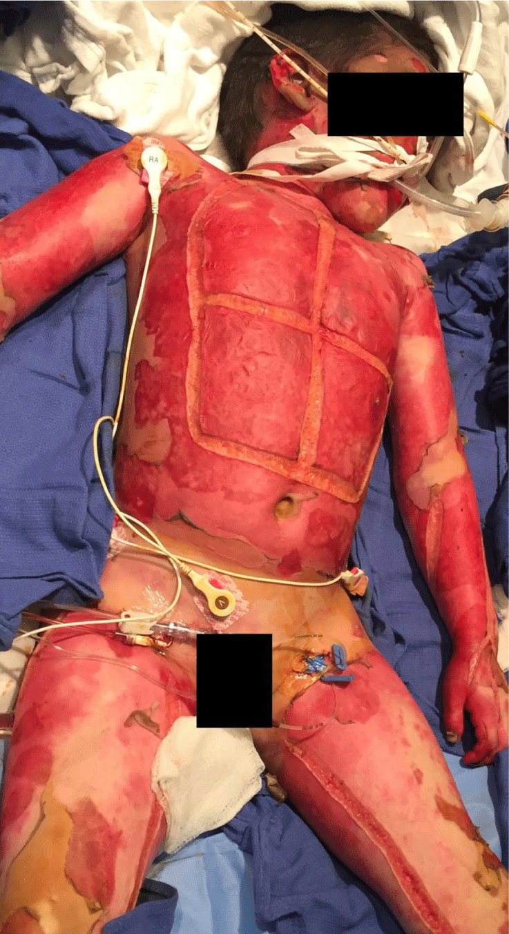
A 5-year-old male was burned over 90% of his body, which occurred during a house fire. The patient and his sister were rescued from the fire and brought to the regional burn center for emergency treatment. The patient was burned all over his body, sparing only his back and groin (Figure 1). During his initial resuscitation, femoral central venous line and arterial lines were placed for monitoring and infusions with the patient under sedation. The patient was gravely ill from the large burn, requiring escarotomies on multiple sites and he developed acute renal failure that required Continuous Renal Replacement Therapy (CRRT) despite adequate fluid resuscitation. To facilitate CRRT, a 12 French acute hemodialysis catheter was placed in the left femoral vein, while the existing central line remained in the right femoral vein. No other sites were available for cannulation. This arrangement left the patient with both the artery and vein cannulated in the same groin, which was not ideal. The typical practice at our regional burn center is to switch venous catheter sites every 7-10 days to minimize the risk for Catheter Related Blood Stream Infection (CRBSI). When the central venous catheter's function started to fail and with no other sites available for catheter placement, a discussion ensued over what course of action to follow. It was decided to prepare the skin near the left clavicle as a site for an initial skin graft, so a central line could be placed in the subclavian vein. One week after excision and debridement, a meshed partial thickness skin graft was taken from the right groin and placed over the left periclavicular area (Figure 2). The graft was trapezoidal in shape, approximately 7 centimetres long at the base and 4 centimetres wide at its maximum. One week later approximately 60% of the graft had healed with the remainder being absorbed >. Despite the approximate 40% of graft loss, the healed skin graft area was large enough for central line placement. Using the seldinger technique, a 5 French double lumen central venous catheter was placed into the left subclavian vein and its position was confirmed by radiography. The patient received sedation for the central line placement. The catheter was used without problems or infection. The process was replicated over the right clavicle and we were able to successfully place a central line into the right subclavian vein through intact, autografted skin. Of note, the Doppler ultrasound monitoring of the venous system, revealed a non-occlusive thrombus in the right femoral vein, which resolved once the catheter was removed.
Discussion
All patients, but children in particular, are at high risk of multisystem organ failure after large 2nd and 3rd degree burns. Central venous catheters are of vital importance in patients with large body surface area burns in order to provide intense supportive carethatis needed to sustain vital organ function. These catheters allow for medication infusion, monitoring of venous pressure, and as in this case, for CRRT. However, use of central venous catheters place patients at risk for CRBSI, a costly [3-6] complication that can lead to increase in length of hospitalization [3-5] and thrombosis [7]. Patients with large body surface area burns are at increased risk from all infections, notably pneumonia, urinary tract infections and CRBSI [1-8]. The increased rate of CRBSI is due to two proposed mechanisms; hematologic seeding of catheters during burn debridements and placement of catheters through or near burns [9]. Due to this high rate of CRBSI many centers, including our burn center, prefer to routinely change central lines. This practice is supported by some studies that suggest changing catheters every 7-10 days decreases infection rate [10,11]. Re-wiring of central catheters is performed by some centers to avoid using additional central venous sites. However, in pediatric burn patients there is an increased rate of CRBSI as compared to selecting a new site [12]. Thus, it is our preferred method to place new catheters at different sites.
Many patients with large body surface area burns have very few sites for placement of central venous catheters. Due to the loss of skin barriers, there is an increased risk of infection during placement and it is difficult to secure the new line to non-intact skin. Further, studies show that placement of catheters within 5 cm of intact skin had a decreased infection rate [13,14]. For these reasons we prefer to place catheters through intact skin. However, we did not have the luxury of placing our catheters through intact skin in this patient due to the extensive nature of the injury, causing us to struggle to find sites to rotate the catheters. After discussions with the surgical team; it was decided to attempt skin grafting over the site of insertion of the subclavian vein. From a surgical perspective, due to the limited amount of non-burned skin, sites with essential function, such as the hands and face, would take priority over the grafting of the periclavicular are. However, given sufficient graft tissue, the team agreed to place a small meshed autologous skin graft over the clavicle due to severity of the multisystem organ failure and need for adequate central venous access.
Allograft is cadaver skin that is frequently used in burn patients. The allograft serves to create a barrier that will act like skin, reducing heat and fluid loss as well as reducing the likelihood of infection when autograft is not available or the burn is not ready for autograft placement. Allograft serves as a substitute for the autograft. The use of allograft for the periclavicular site was discussed in this patient given donor sites were limited. However, the body recognizes the allograft as foreign tissue and over the course of 1-3 weeks the allograft will be rejected [15]. There was concern that this rejection would make allograft a less ideal medium to graft for our central line site and for this reason, it was decided to not place allograft over the periclavicular site.
Conclusion
We present the case of a severely burned child with acute renal failure, in who, due to limited venous insertions sites, had early skin grafting over the clavicle. Although there was some graft loss, there was enough healed skin to provide an intact barrier for central line placement. In cases where there are limited sites for vascular access, early skin grafting should be considered to provide alterative sites.
Consent was obtained from the parent of the child in this case report.
References
- National Nosocomial Infections Surveillance System. National Nosocomial Infection Surveillance (NNIS) system report, data summary from January 1992 through June 2004, issued October 2004. Am J Infect Control. 2004; 32: 470-485.
- Friedman BC, Mian MA, Mullins RF, Hassan Z, Shaver JR, Johnston KK. Five-lumen Antibiotic-Impregnated Femoral Central Venous Catheters in Severely Burned Patients: An Investigation of Device Utility and Catheter-Related Bloodstream Infection Rates. Journal of Burn Care and Research. 2015; 36: 493-9.
- Blot SI, Depuydt P, Annemans L, Benoit D, Hoste E, De Waele JJ, et al. Clinical and economic outcomes in critically ill patients with nosocomial catheter-related bloodstream infections. Clin Infect Dis. 2005; 41: 1591-1598.
- Januel J-M, Harbarth S, Allard R, Voirin N, Lepape A, Allaouchiche B, et al. Estimating attributable mortality due to nosocomial infections acquired in intensive care units. Infect Control Hosp Epidemiol. 2010; 31: 388-394.
- Pittet D, Tarara D, Wenzel RP. Nosocomial bloodstream infection in critically ill patients: excess length of stay, extra costs, and attributable mortality. JAMA. 1994; 271: 1598-1601.
- Warren DK, Quadir WW, Hollenbeak CS, Elward AM, Cox MJ, Fraser VJ. Attributable cost of catheter-associated bloodstream infections among intensive care patients in a nonteaching hospital. Crit Care Med. 2006; 34: 2084-2089.
- Ullman AJ, Marsh N, Mihala G, Cooke M, Rickard CM. Complications of Central Venous Access Devices: A Systematic Review. Pediatrics. 2015; 136: 1331-1344.
- Weber J, McManus A, Nursing committee of the International Society for Burn Injuries. Infection control in burn patients. Burns. 2004; 30: A16-A24.
- Goldman DA and Pier GB. Pathogenesis of Infection Related to Intravascular Catheterization. Clin Microbiol Rev. 1993; 6: 176-192
- Sheridan RL, Weber JM, Peterson HF, Tompkins RG. Central Venous Catheter Sepsis with weekly catheter change in Paediatric Burn Patients: an Analysis of 221 catheters. Burns. 1995; 21: 127-129.
- Sheridan RL, Weber JM. Mechanical and infectious complications of central venous cannulation in children: Lessons learned from a 10-year experience placing more than 1000 catheters. J Burn Care Res. 2006; 27: 713-718.
- O'Mara MS, Reed NL, Palmieri TL, Greenhalgh DG, et al. Central Venous Catheter Infections in Burn Patients with Scheduled Catheter Exchange and Replacement. Journal of Surgical Research. 2007; 142: 341-350.
- Ramos GE, Bolgiani AN, Patino O, Prezzavenot G, Guastavino P, Durlach R, et al. Catheter Infection Risk Related to the Distance Between Insertion Site and Burned Area. Journal of Burn Care and Rehabilitation. 2002; 23: 266-271.
- Kealy G, Chang P, Heinle J, Rosenquist M, Lewis R. Prospective comparison of two management strategies of central venous catheters in burn patients. J Trauma. 1995; 38: 344-349.
- Garfein ES, Orgill DP, Pribaz JJ. Clinical applications of tissue engineered constructs. Clin Plast Surg. 2003; 30: 485-498.
Authors submit all Proposals and manuscripts via Electronic Form!


