Effects of Treatment of Varicose Veins on the Occurrence of Complications and to Improve the Quality of Life: Pilot Study?
Goran Batricevic*, Davor Music, Perica Maras, Milosav Smiljanic, Aleksandar Filipovic and Aleksandar Vulic
Clinical Center of Montenegro, Podgorica, Center for Vascular Surgery of Clinic for Surgery
*Address for Correspondence: Goran Batricevic, Clinical Center of Montenegro, Center for Vascular Surgery of Clinic for Surgery, Podgorica, Montenegro, Tel: +382-690-131-323; E-mail: [email protected]
Submitted: 17 February 2018; Approved: 24 February 2018; Published: 26 February 2018
Citation this article: Batricevic G, Music D, Maras P, Smiljanic M, Filipovic A, et al. Effects of Treatment of Varicose Veins on the Occurrence of Complications and to Improve the Quality of Life: Pilot Study. Adv J Vasc Med. 2018; 3(1): 001-005.
Copyright: © 2018 Batricevic G, et al. This is an open access article distributed under the Creative Commons Attribution License, which permits unrestricted use, distribution, and reproduction in any medium, provided the original work is properly cited
Keywords: varicose veins; surgery; EVLA
Download Fulltext PDF
According to statistics, the disorder of venous circulation of the lower extremities is one of the most common diseases in the world. Although treatment of Varicose Veins the lower extremities (VV) advanced with the use of non-invasive-endovenous procedures, the prognosis in the short term is not satisfactory.
The aim of this paper was to examine the results of treatment of VV on the occurrence of complications and to improve the Quality of Life (QoL) 1 year after the Operative (OP) treatment or Endovenous Laser Ablation (EVLA).
Method: Patients with VV that were operated with the classical surgical method were OP group (n-36) and who are treated EVLA were EVLA group (n-36). We examined patients clinically after 1 year of treatment, while their QoL examined with the Venous Clinical Severity Score (VCSS) questionnaire in which clinical features were estimated by 9 characteristics clinical weight of : 0-absence, 1-easy, 2-moderate, 3-difficult.
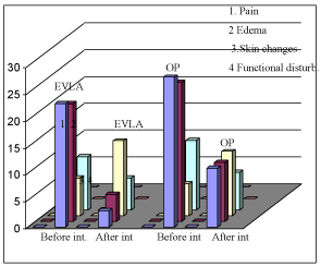
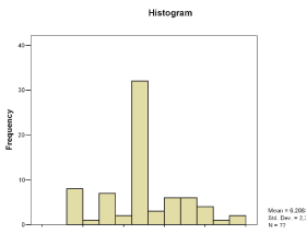
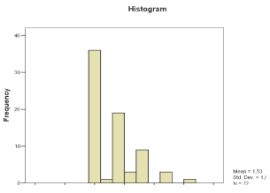
Results: The number of absences from work significantly differed (4.500 ± 5.531 in the EVLA group / 22.944 ± 11.363 in the OP group) (p < 0.000). After interventions, complications were rare, except for hematoma, and there was no difference between groups. QoL after 1 year showed that the VCSS was significantly improved (6.20 ± 2.3 / 1.53 ± 1.88) (p < 0.000). Symptomatology of patients from both groups improved after 1 year of interventions, pain decreased in the OP group by 61%, in the EVLA group by 87% (p < 0.017).
Conclusion: Patients after OP or EVLA treatment of varicose veins had few minor complications and after 1 year of interventions and a better QoL.
Abbreviations
OP: Operative Surgical Ligation of Vein Saphena Magna and Stripping; EVLA: Endovenous Laser Ablation; BMI: Body Mass Index; CEAP classification: Clinical (C), Ethiological (E), Anatomical (A) Pathophysiological (P) [C0-6]; VCSS: Venous Clinical Severity Score; QoL: The Quality of Life; CVI: Chronic Venous Insufficiency; DVT: Deep Vein Thrombosis
Introduction
Venous system may be due to venous wall degeneration, post-thrombotic damage of the valvular apparatus, chronic vein obstruction or disorders of the muscular pump function [1]. All signs and symptoms occurring in CVI are the result. The importance of treating Varicose Veins (vv) is not only for cosmetic reasons, but rather a widespread disease which in the later stage can be difficult. Additionally, vv are disease known for centuries and they are mentioned in Ebers Papirus in ancient Egypt (1580 BC), where it is clearly recommended that they do not work well [2].
The review that collected the results of many English-language studies since 1941 has, as so far shown, a prevalence of Chronic Venous Insufficiency (CVI) that varies, but is most common in Western countries in women > 1% to 40%, and in males > 1% to 17%, depending on the geographic region and risk factors [3].
Dysfunction of the venous hypertension [4]. Histological studies have found in the wall of varicose vein of Vein Saphena Magna (VSM) hypertrophy with increased collagen content and a disorientation of the correct orientation of smooth muscle and elastic fibers [5,6].
Symptoms are mainly, non-specific and depend on the stage in which the disease is located. Patients are most likely to complain about “heavy legs,” pain, swelling, anxiety, night cramps and itching [7]. However, the examination of patients with vv is reduced to four symptoms based on which were made and questionnaires for investigating the success of treatment in pain, edema, changes of the skin and changes in the joints of the feet and as a result disturbed functional ability -Venous Clinical Severity Score (VCSS) questionnaire. These symptoms are the consequence of damage to the veins of the lower extremities.
The weight of the venous disease is classified according to the so-called CEAP classification. This classification is based on certain criteria: 1. Clinical (C), 2. Ethiological (E), 3 Anatomical (A) 4. Pathophysiological (P) [6,7].
Seven clinical categories are defined. C0-C6 which may be symptomatic (S) and asymptomatic (A) [7,8].
The importance of preventing the development of CVI and the timely initiation of treatment for patients, especially in the COs stage, when patients have symptoms despite the lack of visible signs of CVI is that it can normalize venous physiology and delay the further development of the disease [9].
The goals of the treatment of diseased veins are: preventing progression of the disease, alleviation of symptoms, improving, the look of the leg and prevention of complications: thrombophlebitis, Deep Vein Thrombosis (DVT) and lung embolism.
Conventional surgery of varicose veins is based on the physical elimination of the diseased segment, and for the past 20 years the idea has come to that the occlusion of the diseased venous segment (thanks to highly sophisticated duplex scanning, as well as radiofrequency and laser devices) can achieve the same effect, but with less invasive mode [10]. Today, in most developed countries, conventional surgery is slowly replaced by the thermal ablation of the VSM and the Vein Safena Parva (VSP).
The aim of this paper was to examine that the results of treatment of vv on the occurrence of complications and to improve the quality of life 1 year after the Operative (OP) treatment or Endovenous Laser Ablation (EVLA) are better.
Methodology
Patients with vv who were operated with the classical surgical method (OP) were OP group (n-36) and who were treated EVLA were EVLA group (n-36). We examined the patients clinically after 15 days of surgery and after 1 year of treatment for the quality of life with VCSS questionnaire.
The inclusion criteria were: diagnosis of varicose veins prior to the intervention. The exclusion criterion was: patients with other diseases: heart diseases, cancer or some other advanced stage disease. The diagnosis was postulated by clinical inspection and duplex scanning. The severity of the disease was assessed using the VCSS and CEAP classification C0-C6.
Conventional surgery
In all 36 patients, the usual surgical technique was applied. After marking the veins in a standing position, preparation of a safeno-femoral mouth, ligating VSM at the junction, simultaneously with the ligation of lateral branches (crossectomia), then cut in the level of the medial malleolus. After the venotomy a flexible endoluminal catheter is placed, which after the intersection of the vein in the distal part of the ligatures is fixed to the blood vessel in the proximal part. The terminal portion is ligated by the end after the catheter fixation in the groin is accessed by pulling out the entire venous stable (stripping).
Endovenous laser ablation
For the EVLA procedure, we used Biolitek Ceralas TM E laser according to the predicted protocol (Model 980nm / 15w / 200 µm (photo number). Patients were treated with energy (depending on the diameter of the diseased vein) 70-100 J / cm and with a power of 14W continuously at 200 μm fiber diameter across the entire length of the vein.
We used local (tumescent) anesthesia. Puncture a vein was at the predetermined position with the aid of ultrasound, J placing the flexible wire is made hardly visible incision and placing a dilator (sheath). Placing the laser probe (pre-connection to a apparat) through a catheter in which the previously extracted in the dilator position below the saphenous-femoral junction.
Statistical Methods
The statistical analysis were used the descriptive statistics methods. To calculate the difference between the continuous variables of both groups, the hi²-test and t-test were used. The unpaired t-test was used to calculate the effect of improving symptoms before the undertaken intervention and 1 year after interventions in both groups.
Results
The study group consisted of 72 patients who had interventions divided into two groups: Group I-OP (n-36) -patients who operated in Group II-EVLA (n-36) patients who were treated with EVLA Table 1 shows the characteristics of both groups of the patients who did not differ by gender, age, BMI, BMI > 25, CEAP classification-C0-6 and VCSS. A significant difference existed in the number of absent days from work after treatment, so with the patients treated ambulatory EVLA method returns to work faster (group II- 4.500 ± 5.531 days versus group I- 22.944 ± 11.363, p < 0.000).
Discussion
The number of absences from work significantly differed which is a faster recovery and less absence from work, and thus a better socio-economic effect is cited as an important advantage in the EVLA. Many authors emphasize the benefits of outpatient treatment and local anesthesia in EVLA compared to surgery [14].
In our study. After interventions, complications were rare except for haematomas, which were found in both treatment groups and there was no difference between interventions. There were also no complications-Deep Vein Thrombosis (DVT) and Thromboembolism (PE). In other studies, it is also emphasized that complications following intervention are rare, especially the most severe, DVT and PE [13]. Haematomas as the most common complication were recorded in 11 studies, but Carradice et al. reported that significantly less haematomas were in the EVLA group compared to the OP group [14].
We examined patients in terms of their pain, legs edema, changes in the skin (dispigmentation), or possibly causing serious changes and disturbances in daily activities. The results showed that, by observing the total, all patients had a significant improvement in the pain of the legs. Only changes in the skin are worsened, in terms of darker skin, most likely due to insufficient absorption after the intervention hematoma. Other authors also found pain relief [15].
In measuring the quality of life in patients before and after 1 year. From the intervention we used the VCSS questionnaire. There was no difference between the groups, In relation to VCSS prior and after to intervention, the difference was significant. VCSS was significantly lower after 1 year from interventions which means that the quality of life has improved. This result showed, in addition to the assessment of individual symptoms, that interventions significantly improved clinical symptomatology in patients in both groups, but there was no difference between methods, that is, both methods are effective, but that no method was more effective.
In other studies that also used the VCSS scoring system to test the efficacy of EVLA and OP have shown that after interventions, this relationship has significantly improved or decreased VCSS, which means that the quality of life of these patients after interventions has improved. With both methods, without a significant difference between them [15,16]. According to data of some authors improvement of laser technology ,introduction of new apparatus wavelength as 1470 mm which have a higher absorption coefficient of intracellular water of venous wall significantly reducing post innervation pain and significantly less hematoma [17] postoperative pain significantly f > 1% of body surface area) resolve.
Conclusion
OP and EVLA methods of treating varicose veins of the lower extremities are safe and effective, so that patients have had minor complications, following fewer symptoms, especially patients who were in the EVLA group with less pain and edema compared to the operating group and better quality of life.
We need more long terms trials about treatment varicose veins and new technological upgrading to order we resolve the dilemma which the method is superior
The limitation of the study is small numbers of the patients that are studied.
- Gloviczki P, Mozes G. Development and anatomy of the venous system. In: Gloviczki P, editor. Handbook of venous disorders: guidelines of the American Venous Forum. 3rd ed. London: Hodder Arnold; 2009. p. 12-24. https://goo.gl/agfeMm
- Beebe Dimmer JL, Pfeifer JR, Engle JS, Schottenfeld D. The epidemiology of chronic venous insufficiency and varicose veins. Ann Epidemiol. 2005; 15: 175-184. https://goo.gl/MLHE1A
- Ido Weinberg. History of varicose veins.Vascular Medicine. 2011. https://goo.gl/bcuqvT
- Bergan JJ, Schmid Schonbein G, Coleridge Smith P, Nicolaides A, Boisseau M, Eklof B. Chronic venous disease. N Engl J Med. 2006; 355: 488-498. https://goo.gl/hLEcs9
- Travers JP, Brookes CE, Evans J, Baker DM, Kent C, Makin GS, et al. Assessment of wall structure and composition of varicose veins with reference to collagen, elastin and smooth muscle content. Eur J Vasc Endovasc Surg. 1996; 11: 230-237. https://goo.gl/RjoPJf
- Porto LC, Ferreira MA, Costa AM, da Silveira PR. Immunolabeling of type IV collagen, laminin, and alpha-smooth muscle actin cells in the intima of normal and varicose saphenous veins. Angiology. 1998; 49: 391-398. https://goo.gl/WHApVj
- Kistner RL, Eklof B. Classification and etiology of chronic venous disease. In: Gloviczki P, editor. Handbook of venous disorders: guidelines of the American Venous Forum. 3rd ed. London: Hodder Arnold; 2009. p. 37-46. https://goo.gl/5zhW1k
- Gloviczki P, Comerota AJ, Dalsing MC, Eklof BG, Gillespie DL, Gloviczki ML, et al. The care of patients with varicose veins and associated chronic venous diseases: Clinical practice guidelines of the Society for Vascular Surgery and the American Venous Forum. J Vasc Surg. 2011; 53: 2-48. https://goo.gl/iMbHBJ
- Ramelet AA, Perrin M, Kern P, Bounameaux H. Phlebology. 5th ed. Issy les Moulineaux, France: Elsevier Masson; 2008.
- Merchant RF, Pichot O, Closure Study Group. Long-term outcomes of endovenous radiofrequency obliteration of saphenous reflux as a treatment for superficial venous insufficiency. J Vasc Surg. 2005; 42: 502-509. https://goo.gl/SZUGyr
- Maksimovic Zivan; Doric Predrag, Opacic Dragan. Venous insufficiency - innovations. Is conventional surgery outdated? Surgery. 2013; 3: 9-16. https://goo.gl/H5xzML
- Ruchira S W Fernando, Carl Muthu. Adoption of endovenous laser treatment as the primary treatment modality for varicose veins: the Auckland City Hospital experience. N Z Med J. 2014; 127: 43-50. https://goo.gl/Ng3QAZ
- Carroll C, Hummel S, Leaviss J, Ren S, Stevens JW, Everson Hock E, et al. Clinical effectiveness and cost-effectiveness of minimally invasive techniques to manage varicose veins: a systematic review and economic evaluation. Health Technol Assess. 2013; 17: 1-141. https://goo.gl/Fyy2cd
- Carradice D, Mekako AI, Mazari FA, Samuel N, Hatfield J, Chetter IC. Clinical and technical outcomes from a randomized clinical trial of endovenous laser ablation compared with conventional surgery for great saphenous varicose veins. Br J Surg. 2011; 98: 1117-1123. https://goo.gl/x7ebQQ
- Rasmussen LH, Lawaetz M, Bjoern L, Vennits B, Blemings A, Eklof B. Randomized clinical trial comparing endovenous laser ablation, radiofrequency ablation, foam sclerotherapy and surgical stripping for great saphenous varicose veins. Br J Surg. 2011; 98: 1079-1087.
- N S Theivacumar, R Darwood, M J Gough Neovascularisation and Recurrence 2 Years After Varicose Vein Treatment for Sapheno-Femoral and Great Saphenous Vein Reflux: A Comparison of Surgery and Endovenous Laser Ablation. Eur J Vasc Endovasc Surg. 2009; 38: 203-207. https://goo.gl/SgWjgS
- Nedzad Rustempasic, Alemko Cvorak, Alija Agincic. Outcome of Endovenous Laser Ablation of Varicose Veins. Acta Inform Med. 2014; 22: 329-332. https://goo.gl/X7jDL7


Sign up for Article Alerts