Hematological and Pathological Studies of Bacteria Associated with Mobile Phones from Handlers of Diverse Lifestyles in the Rural Community?
MJeffson Avwerosuo Odiete1, Olayiwola JO1* and Abiala MA2
1Department of Biological Sciences, Ajayi Crowther University, Oyo, Oyo State, Nigeria
2Department of Biological Sciences, Mountaintop University, Lagos State, Nigeria
*Address for Correspondence: Olayiwola JO, Department of Biological Sciences, Ajayi Crowther University, Oyo, Oyo State, Nigeria, Tel: +234-810-518-7595; ORCID ID: https://orcid.org/0000-0003-4697-4161; E-mail: olusolajohn97@yahoo.com/ odietejao@gmail.com
Submitted: 29 April 2019; Approved: 10 May 2019; Published: 15 May 2019
Citation this article: Odiete JA, Olayiwola JO, Abiala MA. Hematological and Pathological Studies of Bacteria Associated with Mobile Phones from Handlers of Diverse Lifestyles in the Rural Community. Am J Pharmacol Ther. 2019;3(1): 001-007.
Copyright: © 2019 Odiete JA, et al. This is an open access article distributed under the Creative Commons Attribution License, which permits unrestricted use, distribution, and reproduction in any medium, provided the original work is properly cited
Keywords: Hematology pathological bacteria; Mobile phone; Vehicle
Download Fulltext PDF
Mobile phone has been source of microorganisms that cause diseases of public health concerns. In a study, one-fifth of cellular phones examined were found to harbor pathogenic bacteria indicating that these devices may serve as vehicles of transmission. Swab samples were collected aseptically from the phones of different handlers like motor bike riders, food vendors, meat sellers and nursing mothers. Bacteria isolation and identification were carried out using pour plating technique with distinctive morphological and biochemical characteristics. The pathogenicity of the bacterial isolates was investigated through oral inoculation into albino rats. Eighty-eight (88) bacteria were isolated and selected based on their resistance to antibiotics for pathological study. Loss in weight was observed in some albino rat. Along with reduction in the packed cell volume, hemoglobin but raised white blood cell. Animal inoculated with Bacillus cereus showed meningitis like symptom after the first week of inoculation. Also, there were short and stunted villi; low crystal depth with necrotic debris in the lumen. It has been observed that cell phones may harbor pathogenic bacteria and can subsequently plays role as fomite in the disease transmission. Therefore, the need to educate community phone handlers in the rural area becomes imperative.
Introduction
Mobile phone was established in 1982 in Europe with a view of improving communication [1]. Presently it has become one of the most indispensable devices for professional and social life. It has been discovered that mobile phone harbours different type of organism that are of public health importance [2] through the activities of the handlers. The regular usage and handling of this device, provides an opportunity for the transmission of infections [3-5]. Some of the bacteria reported to be associated with mobile phone include; Klebsiella sp., Salmonella sp., Staphylococcus aureus, Bacillus cereus etc. Muktar et al. [6] reported that the rate of bacterial contamination of mobile phones is very high and that the resistance pattern observed among the bacteria associated is also a huge challenge to the commonly available drugs.
In Nigeria, which is a part of Africa, mobile phone users have increased dramatically. The activities of the phone users play critical role in the contamination of phones and spread of pathogens. Hence, this could enhance transmission of diseases [7]. There has been marked antibiotic resistance in bacteria isolated from mobile phones [8]. Also, gross resistance to Ampicillin, Penicillin, Cloxacillin and Cefuroxime has been documented [9]. Therefore, the work was designed to investigate bacteria associated with mobile phones of different users from rural community and pathogenicity study were subsequently studied.
Materials and Methods
Sample collection
Study was conducted in Oyo State, Nigeria. A total of forty (40) mobile phones were randomly selected from different phone handlers (All the phone users in the selected professions were included in the study). The 10 samples were unbiased distributed among Meat Sellers (MS), Nursing Mothers (NM), Food Sellers (FS) and Commercial Bike Riders (CBR). Four (4) ml of sterile physiological saline was dispensed into a sterile swab stick container and used as a transport medium. The surface of the phones was thoroughly swabbed using swab sticks and then quickly put into its container, sealed and was transported immediately to the laboratory.
Isolation of bacteria
Sample was inoculated into Nutrient Agar and MacConkey Agar using 0.5 ml as the inoculum. The plates were incubated at 37°C for 24-48 hours. The appearance of the colonies on MacConkey agar were monitored and recorded. Distinct colonies were sub-cultured onto Nutrient agar therefore the pure culture were subjected to morphological and biochemical tests according to [10,11] (Table 1).
Maintenance of the experimental albino rat
Male albino rats, rattus norvergicus albinos, (5 weeks old) weighing 120-200 g were purchased from Department of Veterinary Pathology, University of Ibadan, Ibadan. They were housed in the Animal House, Department of Chemical Sciences, Ajayi Crowther University, Oyo and maintained on the formulated rat feed (Protein 21% min, fat 3.5% min, fibre 6.0% max, calcium 0.8%) from Ladokun feeds Ltd, Ibadan, Adequate water was also provided during the study. Animal experiments and housing procedures were performed in accordance to the animal care and ethical rules.
Inoculation of bacterial isolates into experimental albino rat
The pure culture of bacterial isolates was suspended in 0.95% normal saline corresponds to 0.5 McFarland which corresponds to 1.5×10⁸ bacterial suspension/ ml. The animals were divided into six (6) group with two (2) animals in each group and were inoculated orally based on their weight. The weight of the animals that received single dose ranges from (120 g-148 g) while for double dose ranges from (150 g-196 g). Control group was also set up.
Parameters for haematological study
One ml of blood sample was collected from each rat in replicate by inserting a capillary tube into the media canthus of the eye and blood flowed through the capillary tube into an EDTA (50 μl/ ml) tube for haematological analysis. This analysis was carried out according to method of Schalm et al. [12]. Erythrocyte and Total leucocytes count was done using haemocytometer [13]. Haemoglobin concentration and packed cell volume were also determined.
Parameters for histopathological study
The effect of the bacteria isolates on the organ functions were investigated by examining organs like; Liver, Kidney, Spleen, Lungs and Small intestine. The animals were sacrificed using cervical dislocation method. A ventral midline incision was made with scalpel blade from the xiphoid cartilage to the pelvic area. The five organs (liver, kidney, spleen, lungs and small intestine) were harvested and fixed in 10% formalin labeled bottles and histopathological examination was carried out according to method of Adetunji and Anyanwu [13]; Lillie et al. [14].
Result
Determination of bacteria occurrence in the cell phone swabs
Eighty eight (88) bacteria were obtained in this study in which eighty four (84) were Gram positive and four (4) were Gram negative (Table 2). The occurrence of bacteria across the selected mobile phone users (CBR, FV, MS and NM) showed Bacillus spp. (36.3%) had the highest proportion while other bacteria occurred as follows: Staphylococcus sp. (39.7%), Staphylococcus aureus (9%), Corynebacterium xerosis (3.4%), Corynebacterium kutsceri (3.4%), Citrobacter freundii (1.1%), Salmonella sp. (1.1%), Serratia fonticola (1.1%) and Enterobacter sp. (1.1%).
Determination of changes in the body weight of the experimental albino rats
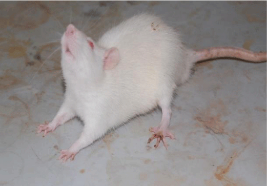
In the animal study, the reduction in body weight was observed in the animal inoculated with Staphylococcus sp. as the weight dropped from 172 g to140 g and 120 g to 81.6 g respectively over the period of three weeks. Similarly, animal challenged with Citrobacter sp. had its weight reduced from 196 g to 144 g over the same period (Figure 1). Interestingly, animals that received dose of Bacillus sp, initially had slight increase in weight in the first week but thereafter, weight reduced. Animal without bacteria inoculation served as control and for the period of experimental procedure the animal remained healthy, the feeding response did not change and the weight of the animal increased as time progresses (Plate 1). Abnormal neck twisting was also observed in the rat inoculated with Bacillus cereus (Plate 2).
Determination of hematological indices in the experimental albino rats
In the haematological study, there was slight reduction in the Pack Cell Volume (PCV), Hemoglobin (HB), White Blood Cell (WBC) but slight increase in the number of platelets in the animal inoculated with Bacillus sp., Bacillus cereus, caused decrease in the PCV, HB, RBC, WBC, Lymphocyte and Neutrophil but slight increase in Monocyte while Eosinophil remain unchanged as observed in the control. Staphylococcus sp. and Salmonella sp. stimulated decrease in PCV, HB and WBC but increase in RBC and Platelet in the rat model (Table 3). There was decrease in Lymphocyte, Monocyte and Eosinophil but increase in Neutrophil in albino rat inoculated with Staphylococcus aureus (Table 4).
Determination of histopathological parameters in the experimental albino rats
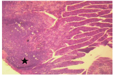
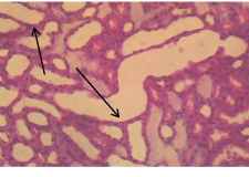
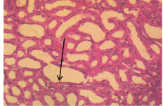
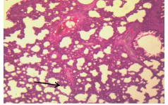
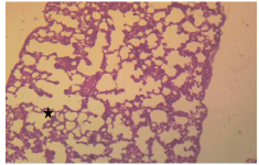
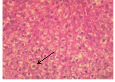
From the histopathological study, the gut-associated lymphoid tissue was large and had prominent germinal centers with moderate numbers of villi which are short and stunted, low cryptal depth with necrotic debris in the lumen as demonstrated by Bacillus cereus (Plates 3&4). Multiple foci of moderate flattening of tubular epithelium with the affected tubules appearing dilated in the kidney and closely-packed hepatic plates (Plates 5&6). There was appearance of large and discrete lymphoid follicles with moth-eaten appearance of germinal center of the spleen in the albino rat inoculated with Citrobacter sp. There was over-distention (emphysema) of alveoli due to some foci thickening of alveolar wall of the Lung (Plates 7&8). However, albino Rat challenged with Staphylococcus sp. showed a few foci of hepatocellular necrosis of the liver. There are a few foci of mild thinning of hepatic cords around the central veins resulting in dilated sinusoids in the liver of the albino rat inoculated with Bacillus sp. (Plates 9&10). The bacteria inoculation result in associated large and discreet lymphoid follicle with foci of pigment-laden macrophages in the spleen (Plates 11&12).
Discussion
Mobile phone is a potential source of disease transmission which plays the role of fomites. Pathogenic organisms have isolated from the swabbed samples of mobile phones as observed in our study which is similar to Famurewa and David, [15] that reported cell phone as a medium of transmission of pathogens.
Girma et al. [16] reported contamination of cell phone and associated potential hazards. The findings in this study showed that some of the pathogens present on the surface of the mobile phones were potential hazard if the organisms find their ways into the susceptible host which could be animal or human especially whenever adequate hygiene is lacking.
In the animal study, it was observed that there was reduction in the weight of the animal after the third week of inoculation which corresponds to the study conducted by Adetunji and Anyanwu [13]. There was significant reduction in the level of PCV, RBC and Hb in the rat inoculated with Bacillus sp., and Citrobacter sp. which is similar to the study of Anubama et al. [17] and Adetunji and Anyanwu [13]. The reduction of PCV, erythrocyte and Hb may be attributed to more than one factor, which is the failure to supply the blood circulation with cells from haem hepatic tissues, since the liver has an important role in the regeneration of erythrocyte or possible destructive effect on erythrocyte pathogenesis of the bacteria inoculated. There was a significant decrease in lymphocyte in all the animals inoculated with bacteria however, there was increase in the humoral immune response like neutrophil eosinophil and monocytes as observed in this study especially in rat inoculated with Citrobacter sp. and Staphylococcus sp. respectively. This is similar to report of Adetunji and Anyanwu [13].
Also, there were numerous closely-packed villi, gut-associated lymphoid tissue is large and prominent germinal centres which could account for reduced surface area thereby reducing rate re-absorption of digested food. Some pathology revealed that wall of small intestine possesses moderate numbers of villi which are short and stunted. Necrotic debris in the lumen was observed in the study especially Bacillus cereus that showed sign of meningitis which has been sometime reported by Stevens et al. [18]. It was also observed that affected tubules of the kidney appeared dilated.
It was observed that alveoli of the lung become over-distended (emphysema) due to some foci of thickening of alveolar wall. Also, bronchioles and alveoli become marked with thickening of alveolar interstitium through inoculation with Citrobacter sp, Staphylococcus sp. similar to the result of Sherein et al. [19]. The hepatic plates of the liver become closely-packed together and few foci of hepatocellular necrosis due to inoculated Citrobacter sp and Staphylococcus sp. which is similar to the result obtained by Abdul-kareem [20] who inoculated rat with Enterobacter cloacae. It was also observed that there were large and discreet lymphoid follicles of the spleen which appeared moth-eaten at the centre revealing due to Citrobacter sp. and Staphylococcus sp.
Conclusion
This study showed that mobile phones may serve as a vehicle for the transmission of pathogenic organisms thereby resulting in disease condition. It can also be deduced that some of the bacteria associated with phone could have implication in condition that may be anaemic among infectious pathogens. Therefore, there is need to mount up an intervention programme to educate the people on impact of phone as a form of fomite and the hygienic way of handling hand set phones so as to minimize the risk of mobile phones as vehicles for disease transmission.
Acknowledgement
We appreciate all the subjects enrolled in this study for their sincere participation during sample collection. We would like to commend the laboratory assistance provided by Mr I.O. Okunlola and Alice Animashaun for her effort during animal experiment.
Author Contribution
JOO, OJ and MAA designed the research. OJ carried out the laboratory work while JOO supervised the laboratory work. The result was analyzed and interpreted by JOO. All the authors participated in the writing of the manuscript and approved the manuscript.
- Neubauer G, Rooslim KN, Feychting M, Hamnerius Y, Kheifets L, Wiart J, et al. Study on feasibility of epidemiological studies on health effects mobile telephone stations. Journal of Health and Population. 2005; 20: 200-210. https://bit.ly/30bVsep
- Karabay O, Kocoglu E, Tahtaci M. The role of mobile phones in the spread of bacteria associated with nosolomial infections. Journal of Infectious Diseases in Developing Countries. 2007; 1: 72-73.
- Glodblatt JG, Krief I, Klonsky T, Haller D, Milloul V, SixSmith DM, et al. Use of cellular telephones and transmission of pathogens by medical staff in New York and Israel. Infect Control Hosp Epidemiol. 2007; 28: 500-503. https://bit.ly/2HjLnmZ
- Yusha’u M, Bello M, Sule H. Isolation of bacteria and fungi from personal and public cell phones: a case study of Bayero University, Kano (old campus). International Journal of Biomedical and Health Sciences. 2010; 6: 97-102.
- Prakash DP, Pawar SA. Nosocomials Hazards of Doctors Mobile Phones. J Theor and Expe Biol. 2012; 8: 115-121.
- Muktar Gashaw, Daniel Abtew, Zelalem Addis. Prevalence and antimicrobial susceptibility pattern of bacteria isolated from mobile phones of health care professionals working in Gondar town health centers. Hindawi Publishing Corporation ISRN Public Health. 2014. https://bit.ly/2JhfLSg
- Butcher W, Ulaeto D. Contact inactivation of bacteria by household disinfectants. Journal Applied Microbiology. 2005; 99: 79-284. https://bit.ly/2Q3imzN
- Sepehri G, Talebizadeh N, Mirzazadeh A, Mir-shekari T, Sepehri E. Bacterial contamination and resistance to commonly used antimicrobials of healthcare workers' mobile phones in teaching hospitals, Kerman, Iran. American Journal of Applied Science. 2009; 6: 806-810. https://bit.ly/2Hl4flp
- Khan RMK, Malik A. Antibiotic resistance and detection of â-lactamase in bacterial strains of Staphylococci and Escherichia coli isolated from foodstuffs. World J MicrobiolBiotechnol. 2001; 17: 863- 868.
- Cheesbrough M. District laboratory practice in tropical countries, part 2. Cambridge University Press. 2002.
- Criuckshank R, Duguid JP, Marmion DP, Swain P. Medical microbiology 12th edition, Churchhill L Iivingstone, Edinburgh. 2006.
- Schalm OW, Jain NC, Carrol EJ. Veterinary haematological. 3rd edition. Lean and Febiger. Philadelphia. 1975; 421-538.
- Adetunji VO, Anyanwu SC. Clinico-pathological changes in Wistar Rats administered B. thuringiensis isolate from soft cheese Wara. Advances in Biological Research. 2011; 5: 206-214. https://bit.ly/2PXz9Ev
- Lillie RD, Fullmen HM. Histopathologic Technic and Practical Histochemistry. 4th ed. McGraw-Hill Company, New York. 1976.
- Famurewa O, David OM. Cell Phone: a medium of transmission of bacterial pathogens: Marsland Press. World Rural Observations. 2009; 1: 69-72.
- Girma G. Potential health risks with microbial contamination of mobile phones. Global Research Journal of Education. 2015; 3: 246-254.
- Anubama VPH, Honnegowda K, Jayakumar GK, Narayana N, Rajeshwari YB. Effect of doramectin on immune response of rats to SRBC antigen. Indian Vet J. 2001; 78: 779-782.
- Stevens MP, Elam K, Bearman G. Meningitis due to Bacillus cereus: a case report and review of the literature. Can J Infect Dis Med Microbiol. 2012; 23: 16-19. https://bit.ly/2WFVOYe
- Sherein I, Abd El-Moez, Ahmed FY, Samy AA, Aisha RA. Probiotic activity of L. acidophilus against major food-borne pathogens isolated from broiler carcasses. Nature and Science. 2010.
- Abdul-kareem, Salman Al-Yassari. Study the pathogenicity of Enterobacter cloacae in rats that isolated from diarrheatic buffalos calves in Babylon province. AL-Qadisiya Journal of Vet Med Sci. 2015.






Sign up for Article Alerts